. 2003 Nov 12;23(32):10433-44.
doi: 10.1523/JNEUROSCI.23-32-10433.2003.

Localization of a beta-actin messenger ribonucleoprotein complex with zipcode-binding protein modulates the density of dendritic filopodia and filopodial synapses

Affiliations

Localization of a beta-actin messenger ribonucleoprotein complex with zipcode-binding protein modulates the density of dendritic filopodia and filopodial synapses

Taesun Eom et al. J Neurosci. .

Abstract

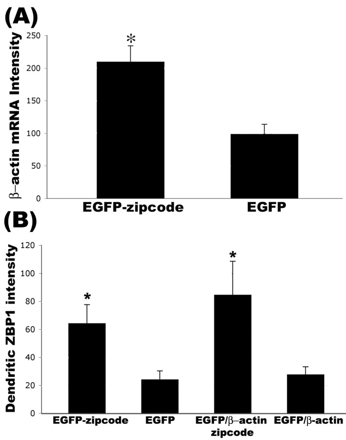
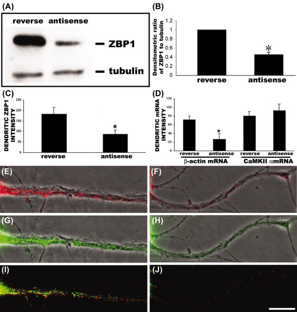
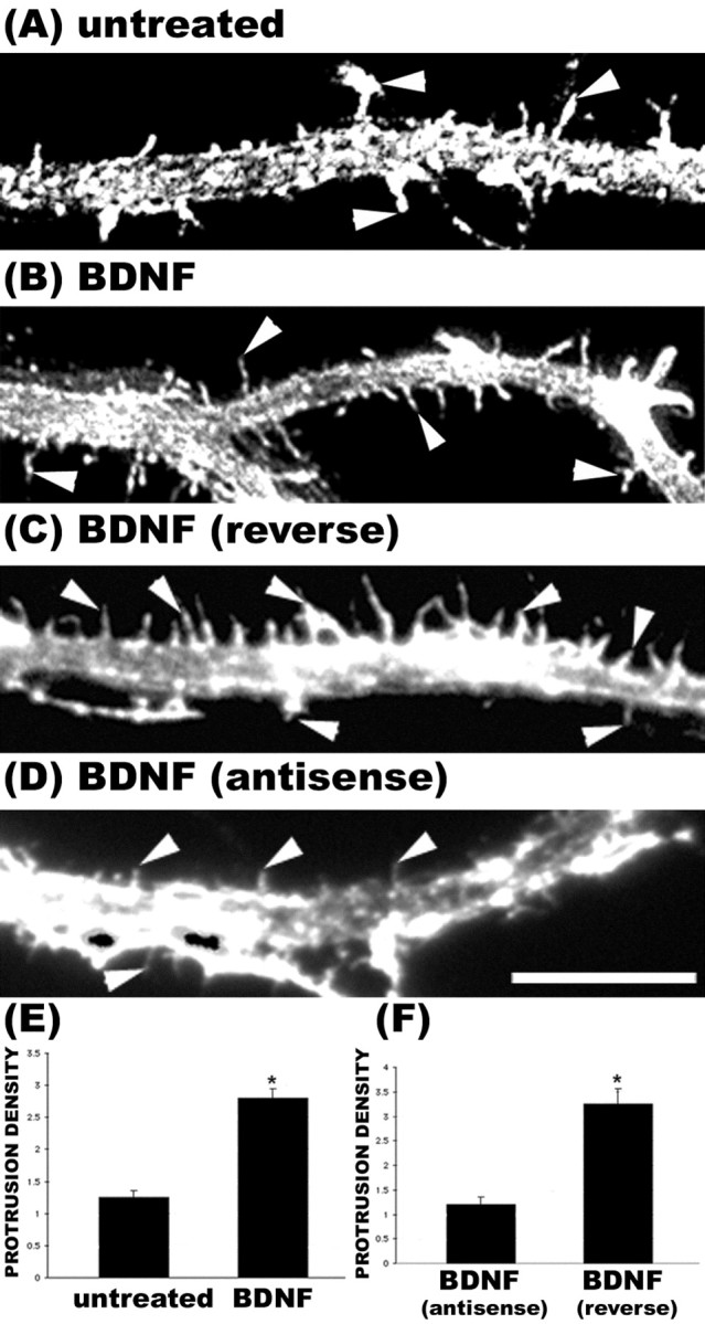
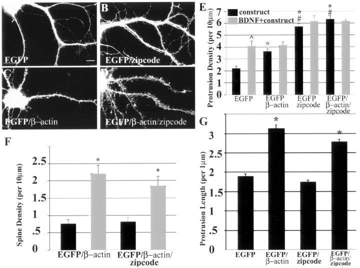
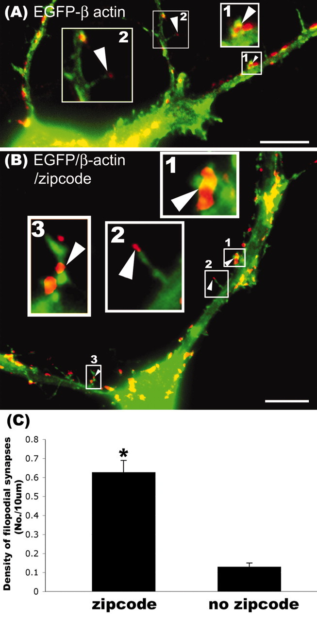
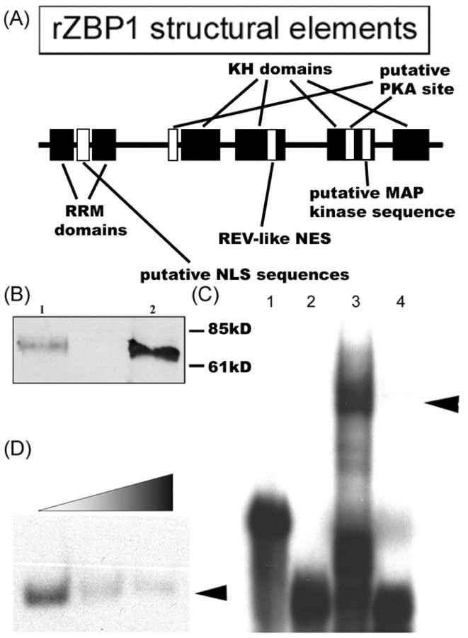
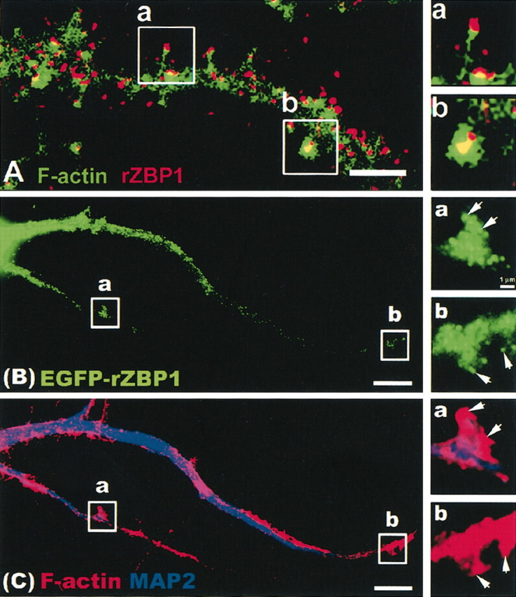
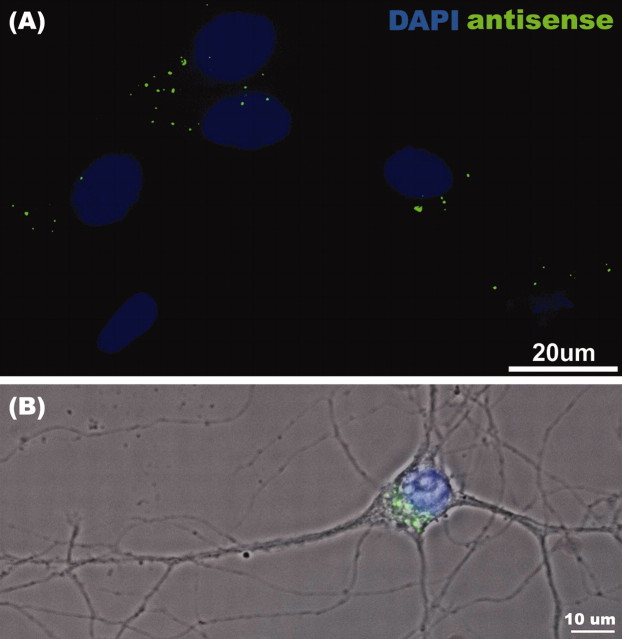
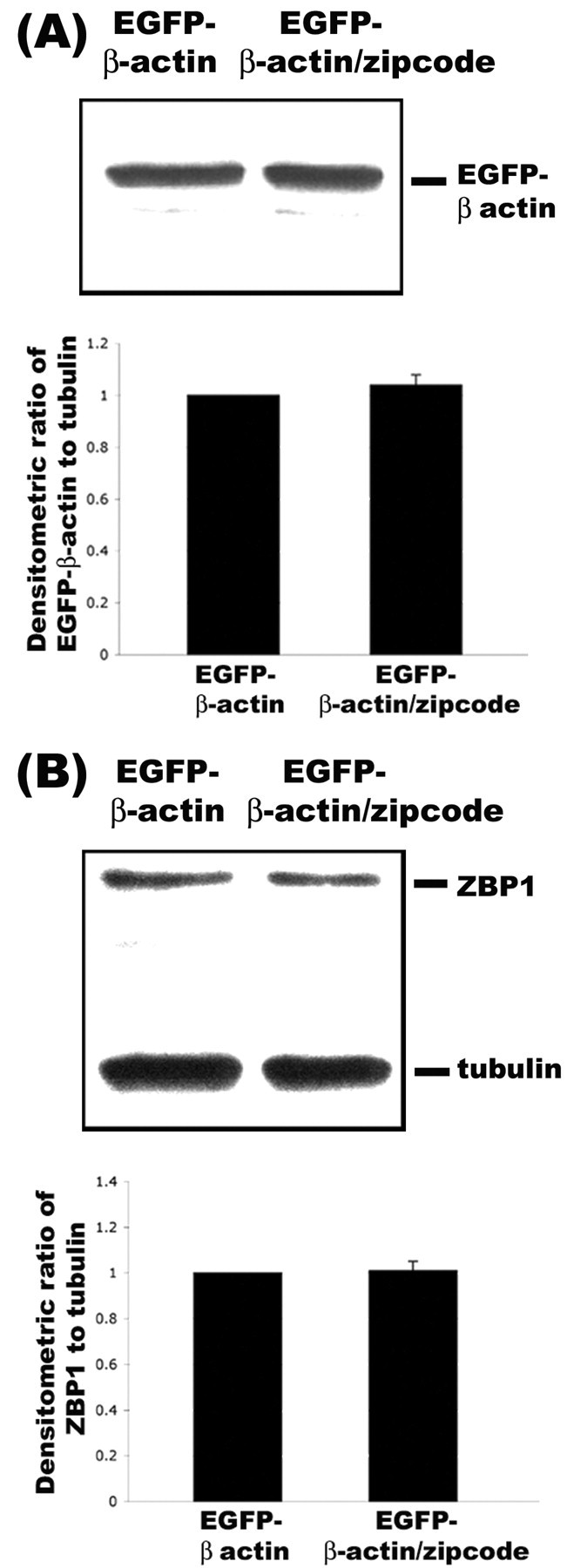
The dendritic transport and local translation of mRNA may be an essential mechanism to regulate synaptic growth and plasticity. We investigated the molecular mechanism and function of beta-actin mRNA localization in dendrites of cultured hippocampal neurons. Previous studies have shown that beta-actin mRNA localization to the leading edge of fibroblasts or the growth cones of developing neurites involved a specific interaction between a zipcode sequence in the 3' untranslated region and the mRNA-binding protein zipcode-binding protein-1 (ZBP1). Here, we show that ZBP1 is required for the localization of beta-actin mRNA to dendrites. Knock-down of ZBP1 using morpholino antisense oligonucleotides reduced dendritic levels of ZBP1 and beta-actin mRNA and impaired growth of dendritic filopodia in response to BDNF treatment. Transfection of an enhanced green fluorescent protein (EGFP)-beta-actin construct, which contained the zipcode, increased the density of dendritic filopodia and filopodial synapses. Transfection of an EGFP construct, also with the zipcode, resulted in recruitment of endogenous ZBP1 and beta-actin mRNA into dendrites and similarly increased the density of dendritic filopodia. However, the beta-actin zipcode did not affect filopodial length or the density of mature spines. These results reveal a novel function for an mRNA localization element and its binding protein in the regulation of dendritic morphology and synaptic growth via dendritic filopodia.


Figures

Figure 8.
Figure 8.
Overexpression of EGFP constructs with the β-actin zipcode resulted in recruitment of endogenous β-actin mRNA and ZBP1. Cultured rat hippocampal neurons were transfected with EGFP, EGFP-zipcode, EGFP-β-actin, and EGFP-β-actin-zipcode. All constructs were expressed for 48 hr. A, Fluorescence in situ hybridization to detect endogenous β-actin mRNA was performed using oligonucleotide probes that excluded the region containing the zipcode. Measurement of average β-actin mRNA levels in dendrites using digital imaging analysis showed an increase in neurons transfected with EGFP-zipcode compared with EGFP alone (*p < 0.05; Student's t test). B, Quantitative immunofluorescence of ZBP1 levels in dendrites showed an increase in both zipcode-containing constructs compared with constructs lacking the zipcode (*p < 0.05; Student's t test; histogram indicates mean values ± SEM).
Figure 3.
Figure 3.
Reduced ZBP1 and β-actin mRNA localization in dendrites after antisense knock-down of rZBP1. A, Western blot analysis with anti-ZBP1 and antitubulin antibodies from extracts of antisense and reverse antisense-treated neurons. Note that the tubulin band is weaker than ZBP because of excessive dilution of antitubulin (1:4000). This prevented oversaturation of the tubulin signal so it could be used for normalization. rZBP1 levels were decreased after 48 hr of antisense treatment, although no changes in tubulin levels were noted. B, Ratio analysis of ZBP1/tubulin band intensities. In antisense-treated cells, the relative intensity of rZBP1 (compared with tubulin) was decreased compared with reverse antisense-treated cells. C, Antisense treatment decreased rZBP1 levels in dendrites, as measured using quantitative immunofluorescence. Cultured rat hippocampal neurons were incubated with either antisense or reverse antisense oligonucleotides to rZBP1 for 48 hr and processed for immunofluorescence. D, Quantitative FISH analysis showed that rZBP1 antisense treatment decreased β-actin mRNA levels in dendrites but not CaMKIIα mRNA levels. E, Example of β-actin mRNA signal in reverse antisense-treated neuron. G, Corresponding signal for ZBP1 immunofluorescence in the same neuron. I, Merge of both β-actin mRNA and ZBP1 showing partial pixel overlap. F, Example of β-actin mRNA signal in rZBP1 antisense-treated neuron. H, Corresponding signal for ZBP1 immunofluorescence in the same neuron. J, Merge of both β-actin mRNA and ZBP1 signal. B, C, D, *, Significant difference in mean values ± SEM; p < 0.05; Student's t test. Scale bar, (E-J) 10 μm.
Figure 5.
Figure 5.
Knock-down of rZBP1 inhibited the growth of dendritic protrusions in response to BDNF stimulation. A, Phalloidin stain of an untreated neuron. B, Phalloidin labeling of a neuron treated with BDNF (25 ng/ml) for 2 hr before fixation. An increase in the density of filopodial-like protrusions was observed (arrowheads). C, Control neurons treated with reverse antisense oligonucleotides to rZBP1 (48 hr) and then stimulated with BDNF (2 hr) also showed an increased density of protrusions (arrowheads). D, In contrast, treatment of neurons with antisense oligonucleotides to rZBP1 resulted in markedly fewer filopodial protrusions after BDNF treatment (arrowheads). E, Quantitation of the density of total F-actin protrusions in untreated and BDNF-treated cultures showed a significant increase after BDNF (p < 0.05). These neurons were not exposed to any morpholino oligonucleotides. F, Quantitation of the density of F-actin protrusions in BDNF-treated cultures exposed to reverse antisense or antisense morpholino oligonucleotides; note the significantly reduced density in antisense-treated neurons (*p < 0.05; Student's t test; histogram indicates mean value ± SEM). Scale bar, 10 μm.
Figure 6.
Figure 6.
Morphologic analysis of dendritic protrusions in neurons transfected with EGFP constructs. Cultured rat hippocampal neurons were transfected with the following cDNAs: EGFP (A), EGFP-zipcode with the β-actin zipcode in the 3′UTR (B), EGFP-β-actin (C), and EGFP-β-actin-zipcode with the β-actin zipcode in the 3′UTR (D). Neurons were fixed after 48 hr of construct expression. A quantitative morphometric analysis was performed using image analysis software (IP Lab; Scanalytics) to measure mean protrusion density (E), mean protrusion length (G), and mean spine density (F). Spines were defined as a dendritic protrusion with a broad or bulbous morphology in contact with synpasin puncta, which was detected by immunofluorescence. At least 10 neurons per construct were randomly selected, followed by acquisition of GFP fluorescence at constant exposure times. A few dendrites from each neuron were analyzed, and each experiment was repeated using separate cultures. This resulted in several hundred to often thousands of protrusions being quantified for each construct. E, A significant increase in protrusion density was observed after overexpression of EGFP-β-actin without the zipcode when compared with EGFP alone (*p < 0.05; ANOVA). Highly significant increases in protrusion density were observed after overexpression of EGFP-β-actin and EGFP constructs containing the zipcode (*p < 0.001; ANOVA). Both zipcode-containing constructs resulted in highly significant increases in protrusion density when compared with β-actin lacking the zipcode (#p < 0.01; ANOVA). A significant increase in protrusion density was observed in EGFP-transfected cells after BDNF stimulation (^p < 0.001). Zipcode-containing constructs seemed to occlude any additional increase in protrusion density by BDNF treatment. F, BDNF treatment significantly increased spine density in neurons transfected with EGFP-β-actin with (*p < 0.01; Student's t test) or without the zipcode (*p < 0.001; Student's t test). G, Overexpression of actin resulted in highly significant increases in the mean length of protrusions, which were not affected by the zipcode (*p < 0.01; Student's t test). Error bars indicate SEM.
Figure 7.
Figure 7.
Overexpression of EGFP-β-actin increased the density of filopodial synapses in a zipcode-dependent manner. Cultured rat hippocampal neurons were transfected with either EGFP-β-actin or EGFP-β-actin-zipcode. Neurons were fixed and stained with antisynapsin antibody (red) as a presynaptic marker. A, EGFP-β-actin (no zipcode): the enlarged insets illustrate and contrast spines with a bulbous or headed morphology (box 1, arrowhead) with those having a long, thin filopodial morphology (box 2, arrowhead). Both types of spines were in contact with synapsin puncta (red). We referred to the filopodial spines (box 2) as filopodial synapses in the text. B, Expression of EGFP-β-actin-zipcode increased the density of filopodial synapses (insets 2, 3, arrowheads). A bulbous headed spine is also depicted in this dendrite for comparison (box 1, arrowhead). C, Quantitation of the average density of filopodial synapses showed that the zipcode significantly increased their density after transfection of EGFP-β-actin (*p < 0.05; Student's t test). Scale bar, 10 μm.
Figure 1.
Figure 1.
Molecular analysis of rZBP1. rZBP1 was cloned from embryonic rat hippocampus using PCR (see Materials and Methods), and its sequence has been entered into GenBank (accession number AF541940). A, rZBP1 structural elements. B, Western blot analysis of recombinant rZBP1 (lane 1) and endogenous rZBP1 from E18 rat hippocampus (lane 2) showed a single band at ∼69 kDa using a rabbit polyclonal antibody to cZBP1. C, An mRNA protein complex was indicated between the β-actin zipcode and rat brain protein extracts using EMSA (lane 3), which was not observed in the absence of extract (lane 1). The control probe migrated to the bottom of the gel in the presence (lane 4) or absence of protein extract (lane 2). D, Formation of an mRNA protein complex between zipcode and recombinant rZBP1 was significantly inhibited by increasing amounts of unlabeled probes.
Figure 2.
Figure 2.
Dendritic localization of endogenous rZBP1 and EGFP-rZBP1. A, Immunofluorescence detection of ZBP1 (red) using a polyclonal antibody. F-actin was detected in the same neuron using phalloidin (Alexa 488), and images were superimposed. Two segments of interest (a,b) were enlarged (right) to show ZBP1 granules in the tip of a long, thin filopodial protrusion (a) and a spine-like, bulbous or headed protrusion (b). Scale bar, 2.5 μm. B, Transfection of EGFP-rZBP1 in cultured hippocampal neurons followed by immunofluorescence detection (C) of MAP2 (blue) and F-actin using TRITC-labeled phalloidin (red). B, C, Two dendritic segments (a,b) are enlarged (right) showing the EGFP-rZBP1 signal (B) and MAP2 and phalloidin signals (C). Arrows denote localization of EGFP-rZBP1 granules in F-actin-rich structures that protrude from the dendritic shaft. Scale bar, 10 μm.
Figure 4.
Figure 4.
Efficient entry of morpholino antisense oligonucleotides into cultured hippocampal neurons. Cultured neurons were transfected with morpholino oligonucleotides (as described in Materials and Methods) with the inclusion of an FITC-conjugated tracer oligonucleotide. A, Cells were fixed and stained with 4′,6′-diamidino-2-phenylindole (DAPI) to reveal nuclei; note five of six cells with FITC labeling in the cytoplasm. B, Higher magnification reveals the presence of FITC-morpholino antisense oligonucleotide in the perinuclear cytoplasm by overlay of the fluorescence and phase images. DAPI-stained nucleus is shown in blue.
Figure 9.
Figure 9.
Expression levels of EGFP-β-actin were not dependent on the presence of zipcode. A, EGFP-β-actin constructs without the zipcode (lane 1) and with the zipcode (lane 2) were transfected into human embryonic kidney 293 cells and processed by Western blot analyses with antibody to EGFP. Quantitative analysis of EGFP levels relative to tubulin using densitometry did not reveal any differences in their levels (bottom). B, From the same lysates, antibodies to ZBP1 were also used for Western blot analysis. No changes in ZBP1 levels were observed relative to tubulin as a loading control (bottom).
None

References

    1. Bassell GJ, Zhang HL, Byrd AL, Femino AM, Singer RH, Taneja KL, Lifshitz LM, Herman IM, Kosik KS ( 1998) Sorting of beta actin mRNA and protein to neurites and growth cones in culture. J Neurosci 18: 251-265. - PMC - PubMed
    1. Bassell GJ, Oleynikov Y, Singer RH ( 1999) The travels of mRNAs through all cells large and small. FASEB J 13: 447-454. - PubMed
    1. Blichenberg A, Schwanke B, Rehbein M, Garner CC, Richter D, Kindler S ( 1999) Identification of a cis-acting dendritic targeting element in MAP2 mRNAs. J Neurosci 19: 8818-8829. - PMC - PubMed
    1. Blichenberg A, Rehbein M, Muller R, Garner CC, Richter D, Kindler S ( 2001) Identification of a cis-acting dendritic targeting element in the mRNA encoding the alpha subunit of CaMKII. Eur J Neurosci 13: 1-9. - PubMed
    1. Burgin KE, Waxham MN, Rickling S, Westgate SA, Mobley WC, Kelly PT ( 1990) In situ hybridization histochemistry of Ca2+/Calmodulin-dependent protein kinase in developing rat brain. J Neurosci 10: 1788-1798. - PMC - PubMed

Publication types

MeSH terms