. 2003 Nov 25;100(24):14000-5.
doi: 10.1073/pnas.2334584100. Epub 2003 Nov 12.

The laminin receptor modulates granulocyte-macrophage colony-stimulating factor receptor complex formation and modulates its signaling

Affiliations

The laminin receptor modulates granulocyte-macrophage colony-stimulating factor receptor complex formation and modulates its signaling

Jian Chen et al. Proc Natl Acad Sci U S A. .

Abstract

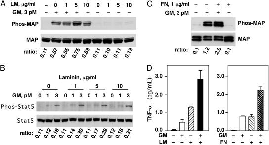
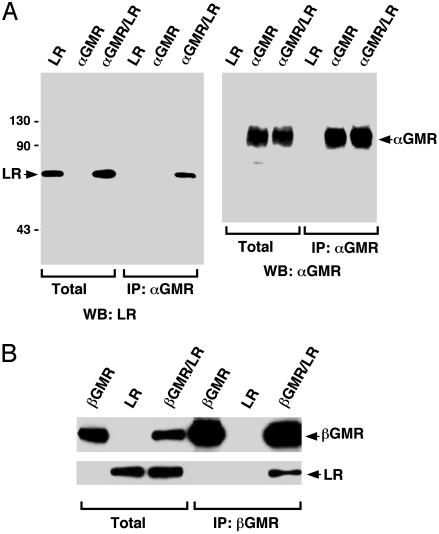
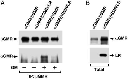
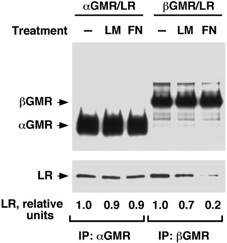
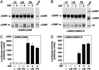
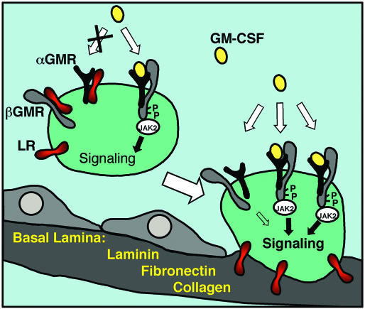
Basement membrane matrix proteins are known to up-regulate granulocyte-macrophage colony-stimulating factor (GM-CSF) signaling in neutrophils and mononuclear phagocytes, but the mechanisms involved are poorly understood. We used the intracellular portion of the alpha subunit of the GM-CSF receptor (alphaGMR) to search for interacting proteins and identified the 67-kDa laminin receptor (LR), a nonintegrin matrix protein receptor expressed in several types of host defense cells and certain tumors, as a binding partner. LR was found to interact with the beta subunit of the GMR (betaGMR) as well. Whereas GM-CSF functions by engaging the alphaGMR and betaGMR into receptor complexes, LR inhibited GM-CSF-induced receptor complex formation. Laminin and fibronectin binding to LR was found to prevent the binding of betaGMR to LR and relieved the LR inhibition of GMR. These findings provide a mechanistic basis for enhancing host defense cell responsiveness to GM-CSF at transendothelial migration sites while suppressing it in circulation.


Figures

Fig. 3.
Fig. 3.
LM and FN enhance GM-CSF function in U937 cells and neutrophils. (A) U937 cells were serum-starved and stimulated with 3 pM GM-CSF (GM) for 10 min. LM (from murine sarcoma) at the indicated concentrations (0, 1, 5, and 10 μg/ml) was added 30 min before GM-CSF treatment. Phosphorylated MAPK (Upper) was analyzed by Western blotting, and the total protein level of MAPK (Lower) was shown as a control. Phosphorylated MAP to total MAP band intensity ratios are indicated. (B) U937 cells were serum-starved and stimulated with 1 pM or 3 pM GM-CSF (GM) for 10 min. LM (from murine sarcoma) at indicated concentrations was added 30 min before GM-CSF treatment. Phosphorylated Stat5 (Upper) was analyzed by Western blotting, and the total protein level of Stat5 (Lower) is shown as a control. Phosphorylated Stat5 to total Stat5 ratios are indicated. (C) Freshly purified human neutrophils were treated with 1 μg/ml FN (from human plasma) for 30 min before 10-min treatment with 3 pM GM-CSF. Phosphorylated MAPK (Upper) was analyzed by Western blotting, and the total protein level of MAPK (Lower) is shown as a control. Phosphorylated MAP to total MAP ratios are indicated. (D) Human neutrophils were treated with GM-CSF (10 pM), LM (from human placenta, 5 μg/ml), or a combination of both for 16 h. TNF-α in the medium was measured by ELISA (Left). Human neutrophils were treated with GM-CSF (5 pM), FN (from human foreskin fibroblasts, 5 μg/ml), or a combination of both for 16 h, and TNF-α production was measured by ELISA (Right).
Fig. 1.
Fig. 1.
Proteins from U937 bind to the ICD of αGMR. (A) Silver staining of αGMR-ICD/CBD expressed in bacteria. Lane M contains protein molecular weight markers. Lane I contains bacteria total protein lysate input. Lane F is chitin beads flow-through fraction, and lane B contains proteins bound to chitin beads (Upper). The identity of the αGMR-ICD was confirmed by Western blotting by using anti-αGMR Ab C18 (Lower). (B) The chitin beads with the fusion protein αGMR-ICD/CBD were incubated with a total cell extract from U937 cells. After washing and DTT elution, αGMR-ICD and its binding proteins (lane E) were analyzed by SDS/PAGE and silver staining. Control binding proteins (lane C) were those DTT-eluted proteins that bind to native chitin beads. Five proteins bands (bands 1–5) appear to be specific binding proteins to αGMR-ICD, and they were sequenced by MS.
Fig. 2.
Fig. 2.
LR binds to both αGMR and βGMR. (A) The 293T cells were transfected with LR (5 μg) or αGMR (5 μg) or cotransfected with LR (5 μg) and αGMR (5 μg). Total protein lysates and αGMR Ab S20 immunoprecipitated proteins were analyzed by SDS/PAGE followed by Western blotting analysis (WB) by using LR Ab anti-V5-HRP (Left) and αGMR Ab C18 (Right). (B) The 293T cells were transfected with βGMR (5 μg) or LR (5 μg) or cotransfected with βGMR (5 μg) and LR (5 μg). Total protein lysates and βGMR Ab S16-immunoprecipitated (IP) proteins were analyzed by SDS/PAGE followed by Western blotting by using LR Ab anti-V5-HRP (Lower) and βGMR Ab C20 (Upper).
Fig. 4.
Fig. 4.
LR inhibits GMR complex formation. (A) The 293T cells were transfected with αGMR plus βGMR (2.5 μg each) or 2.5 μg of αGMR plus 2.5 μg of βGMR plus 5 μg of LR. After transfection (48 h), cells were harvested and lysed; the lysates were then cleared by centrifugation. The lysates were treated with 0.5 nM GM-CSF for 12 h at 4°C. Immunoprecipitation (IP) was done with βGMR Ab S16. In Western blot, αGMR was detected by C18 Ab and the immunoprecipitated βGMR was detected by Ab C20. (B) Total protein levels of αGMR and LR in lysates used for the immunoprecipitation experiment (A) were detected by C18 Ab and anti-V5-HRP, respectively.
Fig. 5.
Fig. 5.
LM and FN disrupt LR/βGMR complex. The 293T cells were transfected with αGMR plus LR (4 μg each) or βGMR plus LR (4 μg each). After transfection (60 h), cells were harvested and treated with LM (mixture of 5 μg/ml human placenta LM and 5 μg/ml murine sarcoma LM) or FN (mixture of 5 μg/ml plasma FN and 5 μg/ml cellular FN) for6hat room temperature with rocking. Cells were centrifuged and lysed, and LM or FN was added to the lysates to maintain the original concentrations. Immunoprecipitation of cleared lysates was done with αGMR Ab S20 and βGMR Ab S16 at 4°C for 16 h. In Western blotting, αGMR and βGMR were detected with Abs C18 and C20, respectively (Upper). LR was detected with anti-V5-HRP Ab (Lower). Relative ratios of LR to αGMR or βGMR band intensities are indicated.
Fig. 6.
Fig. 6.
LM and FN relieve the LR inhibition on GMR system. (A) The 293T cells were transfected with αGMR (2.5 μg) and βGMR (2.5 μg). Cells were treated with LM (mixture of 2 μg/ml human placenta and 2 μg/ml human cellular) or FN (human plasma, 5 μg/ml) for 10 min followed by 0.5 nM GM-CSF treatment for 1 h at room temperature. Cells were centrifuged and lysed with no further addition of matrix proteins or GM-CSF. Proteins were immunoprecipitated by βGMR Ab S16 and βGMR (Upper) and αGMR associated with βGMR (Lower) were detected with Western blotting. αGMR and βGMR in the total protein lysate are shown (Right). (B) The 293T cells were transfected with αGMR (2.5 μg), βGMR (2.5 μg), and LR (5 μg). Cells were treated as in A. Proteins immunoprecipitated by βGMR Ab S16 and βGMR (Upper) and αGMR associated with βGMR (Lower) were detected with Western blotting. αGMR, βGMR, and LR were detected in the total protein lysate (Right). (C) The 293T cells were transfected with αGMR plus βGMR plus pcDNA3.1/GS empty vector plus 8×GAS-luciferase (2 μg each). Luciferase activity was measured after different treatments as indicated. Human placenta LM and human plasma FN were used at 5 μg/ml, and GM-CSF (GM) was used at 300 pM. (D) The 293T cells were transfected with αGMR plus βGMR plus LR plus 8×GAS-luciferase (2 μg each). Luciferase activity was measured after different treatments as indicated. Human placenta LM and human plasma FN were used at 5 μg/ml, and GM-CSF (GM) concentration was 300 pM.
Fig. 7.
Fig. 7.
Schematic model of the LR modulation of GMR system. In circulating host defense cells that are not attached to the basement membrane, the GMR complex formation is inhibited by the LR through its interaction with αGMR and βGMR. The inhibition results in low responsiveness to the GM-CSF cytokine. In cells attached to the basement membrane, the matrix proteins LM, FN, and collagen bind to the LR and the inhibition of the LR on the GMR system is relieved. The basement membrane attachment results in increased GM-CSF signaling. The mechanism implicates that only cells undergoing transendothelial migration are activated and those nonattaching cells do not get activated even though they might see similar concentration of GM-CSF in the infected local milieu. The exact stoichiometry of the functional GMR complex is not reflected.

Comment in

References

    1. Nathan, C. F. (1989) Blood 73, 301–306. - PubMed
    1. Tourkin, A., Anderson, T., LeRoy, E. C. & Hoffman, S. (1993) Cell Adhes. Commun. 1, 161–176. - PubMed
    1. Young, M. R., Lozano, Y., Djordjevic, A., Devata, S., Matthews, J., Young, M. E. & Wright, M. A. (1993) Int. J. Cancer 53, 667–671. - PubMed
    1. Gasson, J. C. (1991) Blood 77, 1131–1145. - PubMed
    1. Gearing, D. P., King, J. A., Gough, N. M. & Nicola, N. A. (1989) EMBO J. 8, 3667–3676. - PMC - PubMed

Publication types

MeSH terms

Substances