. 2003 Nov 13:4:28.
doi: 10.1186/1471-2202-4-28.

Human neuroepithelial cells express NMDA receptors

Affiliations

Human neuroepithelial cells express NMDA receptors

Christopher D Sharp et al. BMC Neurosci. .

Abstract

L-glutamate, an excitatory neurotransmitter, binds to both ionotropic and metabotropic glutamate receptors. In certain parts of the brain the BBB contains two normally impermeable barriers: 1) cerebral endothelial barrier and 2) cerebral epithelial barrier. Human cerebral endothelial cells express NMDA receptors; however, to date, human cerebral epithelial cells (neuroepithelial cells) have not been shown to express NMDA receptor message or protein. In this study, human hypothalamic sections were examined for NMDA receptors (NMDAR) expression via immunohistochemistry and murine neuroepithelial cell line (V1) were examined for NMDAR via RT-PCR and Western analysis. We found that human cerebral epithelium express protein and cultured mouse neuroepithelial cells express both mRNA and protein for the NMDA receptor. These findings may have important consequences for neuroepithelial responses during excitotoxicity and in disease.


Figures
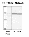
Figure 1
Figure 1
RT-PCR (30 cycles) was performed with primers specific for mouse and human NMDAR1, which revealed that both V1 (mouse neuroepithelial cells) and IHEC (Human cerebral endothelial cells – positive control) express mRNA for NMDAR1. The bands were determined to be ~680 bps and ~670 bps and match the anticipated size of mouse and human NMDAR1 (respectively).
Figure 2
Figure 2
V1 cell protein and rat brain lysate positive control (supplied by Chemicon) were separated on 7.5% denaturing SDS-PAGE gels. Comparison of the control and V1 cell band (~116 kD) present after western analysis demonstrated that V1 cells do express protein for NMDA subunits 1 and 2A/B.
Figure 3
Figure 3
Human hypothalamus sections (A = choriod plexus and B = ependymal cell barrier) were stained for NMDAR1 and demonstrated a uniform and consistent pattern of staining that is associated with the apical side (CSF) with very little staining basally. The magnified section of Figure 3, further illustrates this specific orientation (also pointed out by the arrows). The blood vessels are labeled 'BV' and nuclei are labeled "N".

References

    1. Nicoll RA, Oliet SHR, Malenka RC. NMDA Receptor-Dependent and Metabotropic Glutamate Receptor-Dependent Forms of Long-Term Depression Coexist in CA1 Hippocampal Pyramidal Cells. Neurobiol Learn Mem. 1998;70:62–72. doi: 10.1006/nlme.1998.3838. - DOI - PubMed
    1. Hollmann M, O'Shea-Greenfield A, Rogers SW, Heinemann S. Cloning by functional expression of a member of the glutamate receptor family. Nature. 1989;342:643–648. doi: 10.1038/342643a0. - DOI - PubMed
    1. Bos KD, Slump P. Determination of glutamine and glutamate in plasma of men and women by ion exchange chromatography. Clin Chim Acta. 1985;152:205–211. doi: 10.1016/0009-8981(85)90193-7. - DOI - PubMed
    1. Fonnum F. Glutamate: a neurotransmitter in mammalian brain. J Neurochem. 1984;42:1–11. - PubMed
    1. Coyle JT, Bird SJ, Evans RH, Gulley RL, Nadler JV, Nicklas WJ, et al. Excitatory amino acid neurotoxins: selectivity, specificity, and mechanisms of action. Based on an NRP one-day conference held June 30, 1980. Neurosci Res Program Bull. 1981;19:1–427. - PubMed

MeSH terms

LinkOut - more resources