. 2003 Nov 25;100(24):13875-80.
doi: 10.1073/pnas.2436143100. Epub 2003 Nov 13.

The dual nature of human extracellular superoxide dismutase: one sequence and two structures

Affiliations

The dual nature of human extracellular superoxide dismutase: one sequence and two structures

Steen V Petersen et al. Proc Natl Acad Sci U S A. .

Abstract

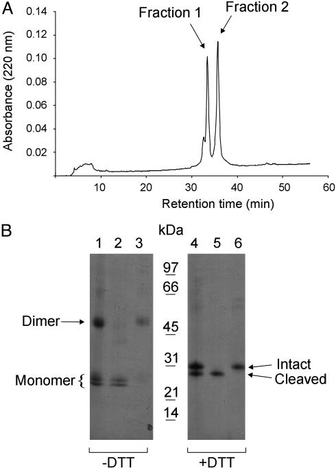
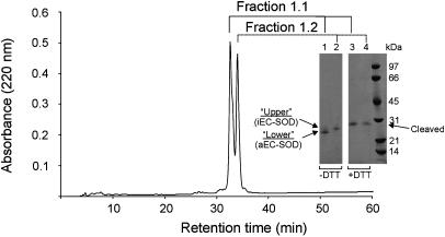
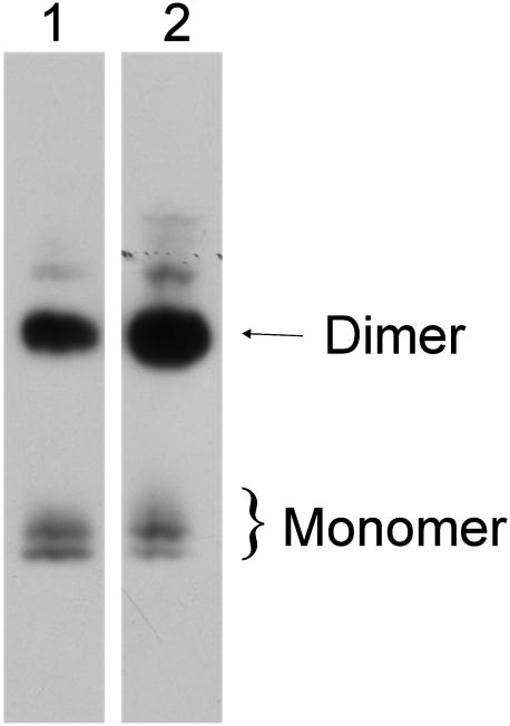
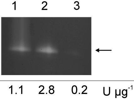
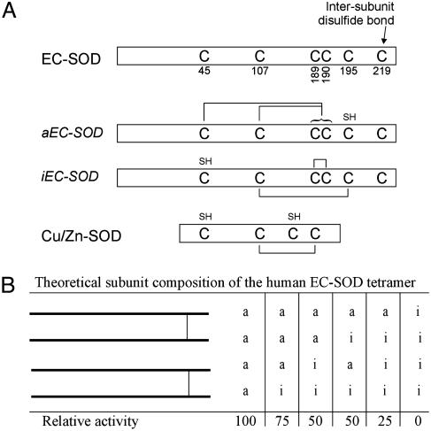
Human extracellular superoxide dismutase (EC-SOD; EC 1.15.1.1) is a scavenger of superoxide anions in the extracellular space. The amino acid sequence is homologous to the intracellular counterpart, Cu/Zn superoxide dismutase (Cu/Zn-SOD), apart from N- and C-terminal extensions. Cu/Zn-SOD is a homodimer containing four cysteine residues within each subunit, and EC-SOD is a tetramer composed of two disulfide-bonded dimers in which each subunit contains six cysteines. The amino acid sequences of all EC-SOD subunits are identical. It is known that Cys-219 is involved in an interchain disulfide. To account for the remaining five cysteine residues we purified human EC-SOD and determined the disulfide bridge pattern. The results show that human EC-SOD exists in two forms, each with a unique disulfide bridge pattern. One form (active EC-SOD) is enzymatically active and contains a disulfide bridge pattern similar to Cu/Zn-SOD. The other form (inactive EC-SOD) has a different disulfide bridge pattern and is enzymatically inactive. The EC-SOD polypeptide chain apparently folds in two different ways, most likely resulting in different three-dimensional structures. Our study shows that one gene may produce proteins with different disulfide bridge arrangements and, thus, by definition, different primary structures. This observation adds another dimension to the functional annotation of the proteome.


Figures

Fig. 1.
Fig. 1.
Separation of monomeric and dimeric human EC-SOD. (A) Purified EC-SOD was subjected to reversed-phase HPLC by using a C8 column and a shallow gradient of acetonitrile in TFA (0.5% solvent B min–1). The absorption profile at 220 nm is shown, with the two collected peaks indicated. (B) The separated material was analyzed by SDS/PAGE and Coomassie blue staining under nonreducing (–DTT) and reducing (+DTT) conditions as indicated. Lanes 1 and 4 represent purified EC-SOD used for the reversed-phase HPLC separation. Lanes 2 and 5 and lanes 3 and 6 represent the material collected in fractions 1 and 2, respectively. The positions of the disulfide-linked dimer and the monomer (Left) and the intact and cleaved forms under nonreducing conditions (Right) are indicated. A molecular mass marker is shown in the center. The analysis shows that EC-SOD can be separated into monomer and dimer by reversed-phase HPLC.
Fig. 2.
Fig. 2.
Separation of EC-SOD monomers. The material collected in fraction 1 (Fig. 1 A) was alkylated with IAA and rechromatographed by reversed-phase HPLC by using the same conditions as in Fig. 1. The alkylated material separated into two peaks, as detected by absorption at 220 nm. Collected fractions were denoted 1.1 and 1.2 as shown. The separated material was analyzed by SDS/PAGE and Coomassie blue staining under nonreducing (–DTT) and reducing (+DTT) conditions (Inset). Lanes 1 and 3 and lanes 2 and 4 contain material collected in fractions 1.1 and 1.2, respectively. The two closely spaced bands under nonreducing conditions are denoted upper and lower band, as indicated. An arrow indicates the position of cleaved EC-SOD under reducing conditions. A molecular mass marker is included on the right.
Fig. 3.
Fig. 3.
Both EC-SOD monomers are present in human aorta. Purified EC-SOD (lane 1) and human aorta homogenate (lane 2) were analyzed by nonreducing SDS/PAGE and Western blotting. The analysis shows that the two bands corresponding to monomeric EC-SOD are detected both in purified EC-SOD (lane 1) and in the aorta homogenate (lane 2). The two distinct bands are thus present in tissue and are not a result of manipulation during purification.
Fig. 4.
Fig. 4.
Activity stain of separated EC-SOD monomers. The EC-SOD monomers were reconstituted with Cu(II) and Zn(II) and separated by SDS/PAGE. Three micrograms of purified EC-SOD was loaded in lane 1. Approximately 1 μg of recharged monomeric EC-SOD collected in fractions 1.1 and 1.2 (Fig. 2 A) was loaded in lanes 2 and 3, respectively. The arrow indicates the position of both monomeric forms on SDS/PAGE analysis of unboiled EC-SOD. No SOD activity can be detected in lane 3. The specific activity determined by the xanthine oxidase/cytochrome c assay is shown below the gel.
Fig. 5.
Fig. 5.
Tryptic peptide map of EC-SOD monomers. EC-SOD collected in fractions 1.1 and 1.2 (Fig. 2) were digested with trypsin. The generated peptides were separated by reversed-phase HPLC, and the 220-nm traces of aEC-SOD (fraction 1.1) (A) and iEC-SOD (fraction 1.2) (B) are shown. All collected peptides were analyzed by Edman degradation and MALDI-MS. The represented differences between the traces are numbered in bold and are presented in Table 1.
Fig. 6.
Fig. 6.
Schematic showing the different EC-SOD forms. (A) Human EC-SOD (P08294) depicted with cysteine residues indicated (top). The two different forms of EC-SOD are shown in the middle with the determined cysteine connectivity indicated. Underivatized cysteine residues of the native protein are indicated by SH. Human Cu/Zn-SOD (P00441) is aligned at the bottom. (B) The theoretical composition of EC-SOD tetramers and the relative SOD activity of these forms. Two intact disulfide-linked dimers are shown.
Fig. 7.
Fig. 7.
Limited reduction of EC-SOD double peptide. To determine the location of the interpeptide disulfide bond between the two connected peptides detected in iEC-SOD F19 (Table 1), we subjected the double peptide to limited reduction. The peptides were analyzed by MALDI-MS. Without reduction (0 mM DTT; Top), the two connected peptides can be detected individually. This is due to disruption of the disulfide bridge during MALDI analysis. The interpeptide disulfide bond can be reduced by 0.01 mM DTT (Middle). The cysteines involved in this bond are alkylated (+IAA). When fully reduced (1 mM DTT, Bottom), all cysteines of the Leu-187–Arg-202 peptide are alkylated (plus 3× IAA). The singly alkylated Leu-187–Arg-202 generated by 0.01 mM DTT was subjected to ESI tandem MS for further analysis.

References

    1. Fridovich, I. (1995) Annu. Rev. Biochem. 64, 97–112. - PubMed
    1. Zelko, I. N., Mariani, T. J. & Folz, R. J. (2002) Free. Radical Biol. Med. 33, 337–349. - PubMed
    1. Fattman, C. L., Schaefer, L. M. & Oury, T. D. (2003) Free. Radical Biol. Med. 35, 236–256. - PubMed
    1. Marklund, S. L. (1982) Proc. Natl. Acad. Sci. USA 79, 7634–7638. - PMC - PubMed
    1. Marklund, S. L. (1984) Biochem. J. 220, 269–272. - PMC - PubMed

Publication types

LinkOut - more resources