. 2003 Dec;15(12):2792-804.
doi: 10.1105/tpc.016618. Epub 2003 Nov 13.

Tapetum determinant1 is required for cell specialization in the Arabidopsis anther

Affiliations

Tapetum determinant1 is required for cell specialization in the Arabidopsis anther

Shu-Lan Yang et al. Plant Cell. 2003 Dec.

Abstract

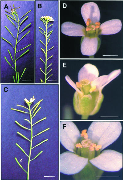
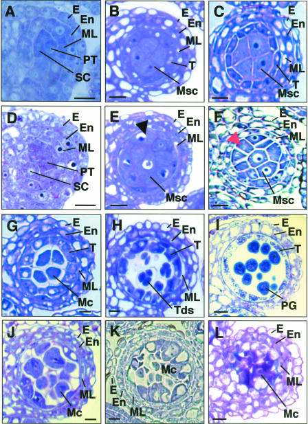
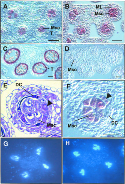
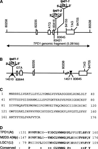
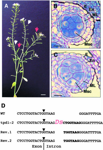
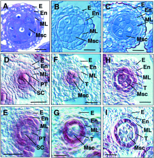
In flowering plants, pollen formation depends on the differentiation and interaction of two cell types in the anther: the reproductive cells, called microsporocytes, and somatic cells that form the tapetum. The microsporocytes generate microspores, whereas the tapetal cells support the development of microspores into mature pollen grains. Despite their importance to plant reproduction, little is known about the underlying genetic mechanisms that regulate the differentiation and interaction of these highly specialized cells in the anther. Here, we report the identification and characterization of a novel tapetum determinant1 (TPD1) gene that is required for the specialization of tapetal cells in the Arabidopsis anther. Analysis of the male-sterile mutant, tpd1, showed that functional interruption of TPD1 caused the precursors of tapetal cells to differentiate and develop into microsporocytes instead of tapetum. As a results, extra microsporocytes were formed and tapetum was absent in developing tpd1 anthers. Molecular cloning of TPD1 revealed that it encodes a small protein of 176 amino acids. In addition, tpd1 was phenotypically similar to excess microsporocytes1/extra sporogenous cells (ems1/exs) single and tpd1 ems1/exs double mutants. These data suggest that the TPD1 product plays an important role in the differentiation of tapetal cells, possibly in coordination with the EMS1/EXS gene product, a Leu-rich repeat receptor protein kinase.


Figures

Figure 1.
Figure 1.
Phenotype of tpd1-1 and a Complemented Mutant. (A) A wild-type plant with fertility indicated by the siliques with normal seed set. (B) A tpd1-1 plant with sterility indicated by the small siliques without seeds. (C) Plant from a transgenic tpd1-1 background line with normal fertility complemented by a wild-type 5.281-kb TPD1 genomic DNA fragment. (D) A wild-type flower, showing the anthers with pollen grains. (E) A tpd1-1 flower, showing the anthers without pollen grains. (F) A complemented tpd1-1 flower, showing the anthers with pollen grains. Bars = 1 cm in (A) to (C) and 1 mm in (D) to (F).
Figure 2.
Figure 2.
Anther Development in tpd1-1 Compared with the Wild Type. The micrographs show one of the four lobes in transverse anther sections. (A) A wild-type anther at stage 4, showing the normal endothecium, middle layer, primary tapetum, and sporogenous cells. (B) and (C) Wild-type anthers at early (B) and late (C) stage 5, showing the well-organized four anther wall cell layers and larger microsporocytes at locule center. (D) A tpd1-1 anther at stage 4, showing the morphologically normal endothecium, middle layer, primary tapetum, and sporogenous cells. (E) and (F) tpd1-1 anthers at early (E) and late (F) stage 5, showing the extra microsporocytes at layer 4, indicated by arrows. (G) A wild-type anther at stage 6, showing the meiocytes detaching from tapetal cells and from each other by depositing callose on the cell walls and the collapsing middle layer. (H) A wild-type anther at stage 7, showing the tetrads. (I) A wild-type anther at stage 11, showing the pollen grains and collapsing tapetal cells. (J) A tpd1-1 anther at stage 6, showing the extra meiocytes and the abnormal middle layer. (K) A tpd1-1 anther at stage 7, showing that no tetrad was formed and that the middle layer was abnormal. (L) A tpd1-1 anther at stage 14, showing the cell debris from the degenerated meiocytes and the highly vacuolated middle layer. E, epidermis; En, endothecium; Mc, meiocyte; ML, middle layer; Msc, microsporocyte; PG, pollen grain; PT, primary tapetum; SC, sporogenous cell; T, tapetum; Tds, tetrads. Bars = 10 μm in (A) to (H) and (J) to (K) and 20 μm in (I) and (L).
Figure 3.
Figure 3.
tpd1-1 Anthers with Extra Microsporocytes and Normal Meiotic Nuclear Divisions but without Tapetum. (A) to (D) and (F) RNA in situ hybridization on transverse sections of wild-type and tpd1 anthers. (A) A wild-type anther at stage 5, showing the specific expression of SDS in microsporocytes. (B) A tpd1 anther at stage 5, showing the expression of SDS in the extra microsporocytes. (C) A wild-type anther at stage 8, showing the specific expression of ATA7 in the tapetal cells. (D) A tpd1 anther at stage 8, showing the absence of the tapetum-specific ATA7 RNA signal. (E) A tpd1 anther at stage 5. The arrow indicates the partially formed microsporocyte cell layer, which is occupied by the tapetum in the wild-type anther. (F) RNA in situ hybridization on a tpd1 anther at stage 5. The arrow indicates the expression signal of SDS in the extra microsporocytes at layer 4. (G) Meiotic nuclear division of a wild-type microsporocyte. (H) Meiotic nuclear division of a tpd1-1 microsporocyte. DC, degenerated cell; ML, middle layer; Msc, microsporocyte; T, tapetum. Bars = 20 μm.
Figure 4.
Figure 4.
Characterization of the TPD1 Gene and tpd1 Mutations. (A) Organization of the TPD1 gene and Ds insertion sites. The rectangular boxes show the exons, and the lines between the exons indicate the positions of the introns. The arrow line indicates the TPD1 genomic fragment used for the complementation of tpd1-1. The numbers indicate the positions of the start and stop nucleotides of the fragments located on BAC clone F6I7. (B) The mutation caused by inversion translocation in the tpd1-1 genome. The solid arrows indicate the recombination sites. The site adjacent to the 3′ end of the Ds element is located in an intergene area, which does not interrupt any predicted gene; the other end is located in the third intron of TPD1 and breaks down TPD1. (C) Sequence of the TPD1 protein with 176 amino acids. (D) Comparison of the TPD1 region at amino acids 131 to 160 with M2D3.4 from liverwort and LGC1 from lily. Boldface letters indicate amino acids that are identical in any two sequences.
Figure 5.
Figure 5.
Phenotype and Characterization of tpd1-2. (A) A tpd1-2 plant with the reversion sectors, showing the revertant siliques (red arrows) adjacent to the small infertile mutant siliques (white arrows). (B) and (C) tpd1-1 and tpd1-2 exhibited similar phenotypes, as demonstrated by the formation of the extra microsporocytes in both tpd1-1 (B) and tpd1-2 (C) anthers at stage 5. (D) The Ds insertion site in the tpd1-2 genome. Boldface letters indicate a typical 8-bp repeat sequence generated by the Ds insertion in the tpd1-2 genome and 7-bp footprint sequences in the revertant genomes. The arrows indicate the splicing site of the third intron. Bars = 1 cm in (A) and 20 μm in (B) and (C).
Figure 6.
Figure 6.
Expression of TPD1. (A) Expression of TPD1 in wild-type seedlings, leaves, and flower buds, as revealed by RNA gel blot hybridization. The bottom gel shows mRNA loading controls. Five micrograms of mRNA was loaded in each lane. (B) to (K) Expression patterns in wild-type flowers, as revealed by RNA in situ hybridization. (B) TPD1 expression in inflorescence apical meristem, floral meristem, and carpel primordia. (C) TPD1 expression in ovule primordia, showing the stronger TPD1 signal in the female archesporial cell. (D) TPD1 expression in archesporial cells in an anther at stage 2. (E) TPD1 expression in primary parietal cells and sporogenous cells in an anther at stage 3. (F) TPD1 expression in secondary parietal cells and sporogenous cells in an anther at stage 4. (G) TPD1 expression in an anther at early stage 5, showing the predominant signal in microsporocytes. (H) TPD1 expression in an anther at late stage 5, showing that the TPD1 RNA signal was predominant in both tapetal cells and microsporocytes. (I) TPD1 expression was reduced greatly in an anther at stage 6. (J) TPD1 expression was not detected in an anther at stage 10. (K) Control section of an anther at stage 5, which was hybridized with the sense TPD1 RNA probe, showing only the background signal compared with the stronger signals on the other sections hybridized with the antisense TPD1 RNA probe. Ar, archesporial cell; CP, carpel primordia; FM, floral meristem; IAM, inflorescence apical meristem; Mc, meiocyte; ML, middle layer; Msc, microsporocyte; Msp, microspore; MW, molecular weight markers; PPC, primary parietal cell; SC, sporogenous cell; SPC, secondary parietal cell; T, tapetum; WT, wild type. Bars = 5 μm in (C) and 20 μm in all other panels.
Figure 7.
Figure 7.
Comparison of tpd1-1 and ems1/exs1-2 and Their Expression Patterns in Anthers by in Situ Hybridization. (A) to (C) One of the four lobes from anther sections of tpd1-1 (A), ems1/exs1-2 (B), and tpd1-1 ems1/exs1-2 (C) at stage 5, showing that they exhibited similar phenotypes, as demonstrated by the formation of extra microsporocytes. (D) and (E) Wild-type anthers at late stage 4. (D) Predominant TPD1 expression in sporogenous cells. (E) Predominant EMS1/EXS expression in the primary tapetum. (F) and (G) Wild-type anthers at early stage 5. (F) TPD1 RNA was detected predominantly in microsporocytes. (G) EMS1 RNA was found mostly in tapetal cells. (H) and (I) Wild-type anthers at late stage 5. (H) TPD1 was expressed mostly in the tapetum and microsporocytes. (I) EMS1 was expressed predominantly in the tapetum. E, epidermis; En, endothecium; ML, middle layer; Msc, microsporocyte; PT, primary tapetum; SC, sporogenous cell; T, tapetum. Bars = 20 μm.

Similar articles

Cited by

References

    1. Azumi, Y., Liu, D., Zhao, D.Z., Li, W., Wang, G.F., Hu, Y., and Ma, H. (2002). Homolog interaction during meiotic prophase I in Arabidopsis requires the SOLA DANCERS gene encoding a novel cyclin-like protein. EMBO J. 21, 3081–3095. - PMC - PubMed
    1. Canales, C., Bhatt, A.M., Scott, R., and Dickinson, H. (2002). EXS, a putative LRR receptor kinase, regulates male germline cell number and tapetal identity and promotes seed development in Arabidopsis. Curr. Biol. 12, 1718–1727. - PubMed
    1. Dawson, J., Wilson, Z.A., Aarts, M.G.M., Braithwaite, A.F., Briarty, L.G., and Mulligan, N.J. (1993). Microspore and pollen development in six male-sterile mutants of Arabidopsis thaliana. Can. J. Bot. 71, 629–638.
    1. Denis, M., Delourme, R., Gourret, J.P., Mariani, C., and Renard, M. (1993). Expression of engineered nuclear male sterility in Brassica napus. Plant Physiol. 101, 1295–1304. - PMC - PubMed
    1. Goldberg, R.B., Beals, T.P., and Sanders, P.M. (1993). Anther development: Basic principles and practical application. Plant Cell 5, 1217–1229. - PMC - PubMed

Publication types

MeSH terms

Substances

Associated data