. 2003 Nov 14:1:112.
doi: 10.1186/1477-7827-1-112.

LCN6, a novel human epididymal lipocalin

Affiliations

LCN6, a novel human epididymal lipocalin

Katherine G Hamil et al. Reprod Biol Endocrinol. .

Abstract

Background: The lipocalin (LCN) family of structurally conserved hydrophobic ligand binding proteins is represented in all major taxonomic groups from prokaryotes to primates. The importance of lipocalins in reproduction and the similarity to known epididymal lipocalins prompted us to characterize the novel human epididymal LCN6.

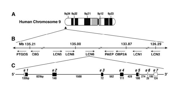
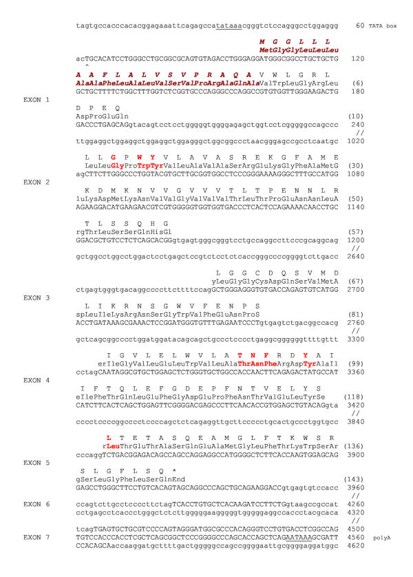
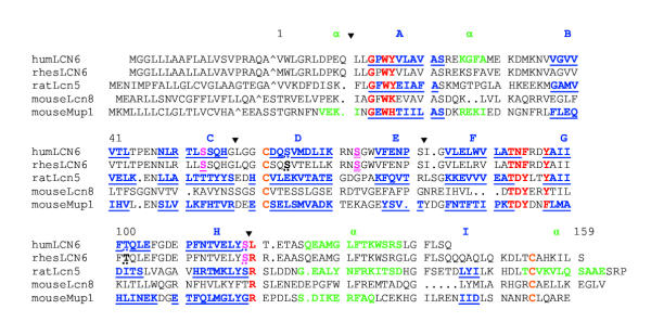
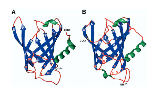
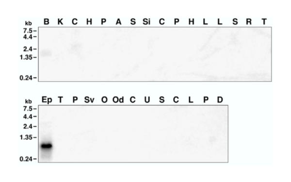
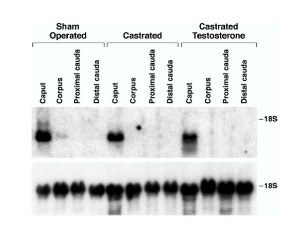
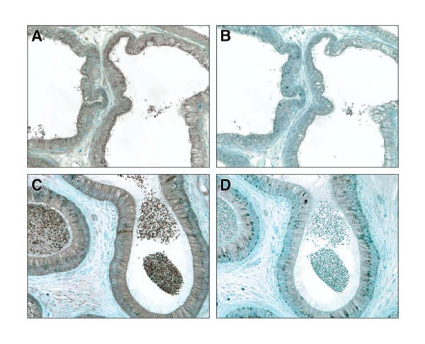
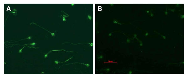
Methods and results: LCN6 cDNA was identified by database analysis in a comprehensive human library sequencing program. Macaca mulatta (rhesus monkey) cDNA was obtained from an epididymis cDNA library and is 93% homologous to the human. The gene is located on chromosome 9q34 adjacent LCN8 and LCN5. LCN6 amino acid sequence is most closely related to LCN5, but the LCN6 beta-barrel structure is best modeled on mouse major urinary protein 1, a pheromone binding protein. Northern blot analysis of RNAs isolated from 25 human tissues revealed predominant expression of a 1.0 kb mRNA in the epididymis. No other transcript was detected except for weak expression of a larger hybridizing mRNA in urinary bladder. Northern hybridization analysis of LCN6 mRNA expression in sham-operated, castrated and testosterone replaced rhesus monkeys suggests mRNA levels are little affected 6 days after castration. Immunohistochemical staining revealed that LCN6 protein is abundant in the caput epithelium and lumen. Immunofluorescent staining of human spermatozoa shows LCN6 located on the head and tail of spermatozoa with the highest concentration of LCN6 on the post-acrosomal region of the head, where it appeared aggregated into large patches.

Conclusions: LCN6 is a novel lipocalin closely related to Lcn5 and Lcn8 and these three genes are likely products of gene duplication events that predate rodent-primate divergence. Predominant expression in the epididymis and location on sperm surface are consistent with a role for LCN6 in male fertility.


Figures

Figure 1
Figure 1
Human LCN6 gene location on chromosome 9q34 A, Ideogram of chromosome 9. Filled triangle near the long arm telomere indicates the location of the lipocalin-rich region containing LCN6. B, Arrangement of lipocalin genes from 126 to 136 megabases from the telomere of the short arm (not to scale). Arrows indicate direction of transcription of each gene. LCN5 and LCN8 indicate uncharacterized genes similar to mouse Lcn5 and 8. Positions were taken from MapView at the The National Center for Biotechnology Information (NCBI) website Human Genome Build 34, Version 1. C, Intron-exon structure of LCN6 gene. Black boxes indicate translated regions and white boxes indicate untranslated regions of exons.
Figure 2
Figure 2
Human chromosomal sequence aligned with LCN6 mRNA and amino acid sequences. The predicted TATA box and polyA addition sites are underlined. Exons are in upper case letters, introns in lower case. The ^ indicates the predicted transcription start site. The amino acids are indicated in single letter and three letter abbreviations. The signal peptide amino acids are brown, bold and italicized. Numbers in parentheses indicate amino acids of the mature protein. The lipocalin signature amino acids are red and bold. The gene sequence was extracted from GenBank accession number AL355987. The human cDNA sequence is available at accession number AF303084 and at EST BU502206.
Figure 3
Figure 3
Amino acid alignment of mature LCN6 and related lipocalins. Lipocalin signature amino acids are in red and cysteines in orange. The predicted casein kinase II phosphorylation sites at S64, T101 and S118 are indicated by dotted underline. Predicted protein kinase A phosphorylation site at Ser73 is magenta and double underlined. Predicted protein kinase C phophorylation sites at S53 and S118 are magenta and single underlined (solid underline for S53 and dotted for S118). Amino acids known to form alpha helices in mouse Mup1 and rat Lcn5 and predicted in LCN6 are green. Beta strands are blue and underlined and labeled A to I corresponding to the β-strands in Fig. 4. The rhesus LCN6 (rhesLCN6) sequence is available at GenBank accession number AF303085. Rat Lcn5 sequence is from accession number X59832 and structural information from 1EPA.pdb and (61). Mouse Lcn8 accession number is NM_033145. Mouse major urinary protein 1 (Mup1) is from M16355. Intron locations in human LCN6 are indicated by the filled triangles (▼). Numbering refers to LCN6 amino acid positions in the mature protein.
Figure 4
Figure 4
Lipocalin models. A, Human LCN6. B, Mouse Mup1 derived from the Protein Data Bank file 1MUP.pdb. Alpha helices are green, beta strands are blue with white letter designations (A-I), unstructured loops are red. The conserved lipocalin signature amino acids are indicated in red. The disulfide bond in MUP1 is indicated as S-S in yellow in the upper left region of the molecule.
Figure 5
Figure 5
LCN6 expression is highly epididymis-specific. Total RNAs were isolated from human urinary bladder, kidney, cerebrum, hypothalamus, pituitary, adrenal, stomach, small intestine, colon, pancreas, heart, lung, liver, spleen, retina, tonsil, epididymis, testis, prostate, seminal vesicle, ovary, oviduct, cervix, uterus, salivary gland and the cell lines CV1, LNCaP, PC3 and DU145 and analyzed by Northern hybridization to 32P-labeled LCN6 full length cDNA.
Figure 6
Figure 6
Androgen regulation of LCN6 mRNA Macaca mulatta were sham operated, castrated, or castrated and androgen replaced with immediate injection of testosterone enanthate. Epididymides were removed 6 days post-castration. Upper Panel Total RNAs (10 μg/lane) from the indicated epididymal regions were analyzed by Northern hybridization to 32P-labeled LCN6 full length cDNA. Lower Panel The same blot was hybridized to 32P-labeled 18S ribosomal RNA cDNA.
Figure 7
Figure 7
LCN6 protein in human efferent ducts and caput. Panels A and B human efferent ducts from an 83 yr old donor. A, LCN6 protein appears brown, distinct from the toluidine blue counterstain. B, A control staining for which antibody was preincubated with 1 mg/ml recombinant human LCN6 protein. Panels C and D human caput. C, LCN6 appears brown. D, Antibody preincubated with 100 μg/ml human LCN6. Photographs were taken using a 20× objective.
Figure 8
Figure 8
Localization of LCN6 on human spermatozoa Panel A, Immunofluorescent staining of spermatozoa with anti-LCN6 Panel B, Control immunofluorescent staining of spermatozoa with anti-LCN6 preincubated with LCN6 protein attached to affinity resin to remove specific antibodies. Original images were taken using a 63× objective.

Similar articles

Cited by

References

    1. Cooper TG. Epididymal proteins and sperm maturation. In: Nieschlag E, Habenicht UF, editor. In Spermatogenesis-Fertilization-Contraception. Berlin/New York:Springer-Verlag; 1992. pp. 285–318.
    1. Cuasnicú PS, Cohen DJ, Ellerman DA, Buso D, DaRos VG, Morgenfeld MM. Changes in specific sperm proteins during epididymal maturation. In: Robaire B, Hinton BT, editor. In The Epididymis From Molecules to Clinical Practice. New York: Kluwer Academic/Plenum; 2002. pp. 389–403.
    1. Dacheux JL, Gatti JL, Dacheux F. Contribution of epididymal secretory proteins for spermatozoa maturation. Microsc Res Tech. 2003;61:7–17. doi: 10.1002/jemt.10312. - DOI - PubMed
    1. Rankin TL, Tsuruta KJ, Holland MK, Griswold MD, Orgebin-Crist MC. Isolation, immunolocalization and sperm-association of three proteins of 18, 25 and 29 kilodaltons secreted by the mouse epididymis. Biol Reprod. 1992;46:747–766. - PubMed
    1. Gerena RL, Irikura D, Eguchi N, Urade Y, Killian GJ. Immunocytochemical localization of lipocalin-type prostaglandin D synthase in the bull testis and epididymis and on ejaculated sperm. Biol Reprod. 2000;62:547–556. - PubMed

Publication types

MeSH terms