

Common arterial trunk in the fetus: characteristics, associations, and outcome in a multicentre series of 23 cases
- PMID: 14617557
- PMCID: PMC1767971
- DOI: 10.1136/heart.89.12.1437
Common arterial trunk in the fetus: characteristics, associations, and outcome in a multicentre series of 23 cases
Abstract
Objective: To assess the accuracy of prenatal diagnosis, the incidence of extracardiac and chromosomal anomalies, and the perinatal outcome in a population of fetuses with common arterial trunk (CAT).
Design: Observational study of 23 fetuses from three referral centres with a confirmed diagnosis of CAT. All underwent fetal echocardiography, detailed anatomical scanning, and karyotyping. In 19 cases, FISH analysis was done to detect 22q11 microdeletion. The following variables were evaluated: gestational age at diagnosis, anatomical variants of the CAT, presence of extracardiac and chromosomal anomalies, pregnancy, and fetal-neonatal outcome. Necropsy reports and postnatal files were available for confirmation of the prenatal diagnosis in all cases.
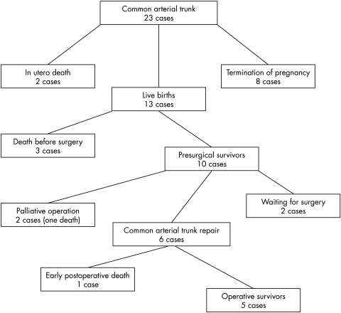
Results: The prenatal diagnosis proved correct in 23 of 24 cases, the last being pulmonary atresia with ventricular septal defect (PAVSD). A second cardiovascular anomaly was present in eight cases (34.8%); extracardiac anomalies were found in 10 (43.4%). FISH analysis showed 22q11 microdeletion in six of 19 cases (31.6%). Outcomes were as follows: eight terminations of pregnancy (34.8%), two intrauterine deaths (8.7%), five postnatal deaths (before or after surgery) (21.7%); the remaining eight neonates (34.8%) are alive and thriving after surgery (six) or awaiting surgery (two).
Conclusions: CAT can be reliably diagnosed and characterised in prenatal life, although differentiation from PAVSD may be challenging. The association with chromosomal anomalies is consistent (8.7%), but there is a higher risk of 22q11 microdeletion (31.6%), in agreement with postnatal studies. The relatively poor survival rate (34.8%) reflects the high rate of terminations and the unfavourable cardiac anatomy in some cases.
Figures
References
-
- Collett RW, Edwards JE. Persistent truncus arteriosus: a classification according to anatomic types. Surg Clin North Am 1949;29:1245–70. - PubMed
-
- Allan LD, Sharland GK, Milburn A, et al. Prospective diagnosis of 1006 consecutive cases of congenital heart disease in the fetus. J Am Coll Cardiol 1994;23:1452–8. - PubMed
-
- Marasini M, Cordone M, Zampatti C, et al. Prenatal ultrasonic detection of truncus arteriosus with interrupted aortic arch and truncal valve regurgitation. Eur Heart J 1987;8:921–4. - PubMed
-
- Paladini D, Rustico MA, Todros T, et al. Conotruncal anomalies in prenatal life. Ultrasound Obstet Gynecol 1996;8:241–6. - PubMed
-
- Duke C, Sharland GK, Jones AMR, et al. Echocardiographic features and outcome of truncus arteriosus diagnosed during fetal life. Am J Cardiol 2001;88:1379–84. - PubMed
Publication types
MeSH terms
LinkOut - more resources
Full Text Sources
Medical
Miscellaneous