

Review
doi: 10.1136/heart.89.12.1447.
The role of echocardiography in atrial fibrillation and cardioversion
Affiliations
- PMID: 14617563
- PMCID: PMC1767994
- DOI: 10.1136/heart.89.12.1447
Item in Clipboard
Review
The role of echocardiography in atrial fibrillation and cardioversion
Heart.
2003 Dec.
No abstract available
Figures
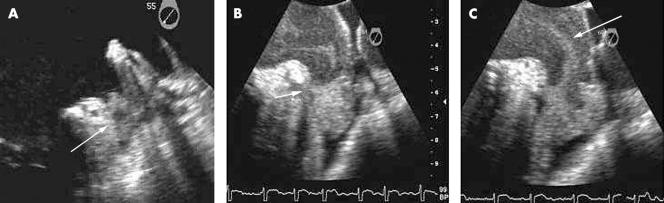
Images of the left atrium and left atrial appendage (LAA) depicting the spectrum of flow stasis and thromboembolic risk from “smoke” through thrombus. (A) LAA thrombus detected in a patient with atrial fibrillation (AF) undergoing transoesophageal echo (TOE) before cardioversion. Note the well formed rounded upper edge of the thrombus (arrow). (B) “Sludge” in the apex of the LAA detected by TOE in a patient with AF. Note the meniscus at the upper edge of the sludge (arrow). (C) Spontaneous echo contrast or “smoke” swirling detected by TOE in the LAA and left atrium of a patient in AF.
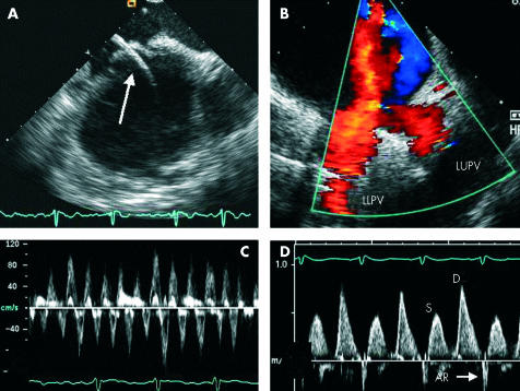
Images acquired from intracardiac echocardiography (ICE). (A) Transeptal puncture with catheter (arrow) passing from right atrium to left atrium. (B) Colour flow mapping showing the confluence of the left upper and lower pulmonary veins (LUPV and LLPV). (C) Pulsed wave Doppler of the LAA showing preservation of velocities in a patient with AF. (D) Pulsed Doppler of the LUPV showing blunting of systolic (S) flow compared to diastolic (D) flow and a narrow atrial systolic reversal (AR) following cardioversion in the same patient.
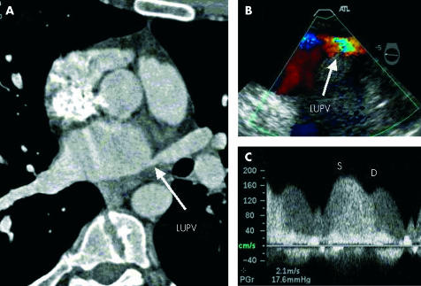
Stenosis of the left upper pulmonary vein (LUPV) following radiofrequency ablation for AF. (A) Narrowing of the LUPV ostium (arrow) on multi-detector computed tomography. (B) Colour Doppler image from TOE demonstrating aliasing of flow in the LUPV. (C) Pulsed wave Doppler of the LUPV demonstrating phasic systolic (S) and diastolic (D) flow with increased velocities (> 1.5 m/s).
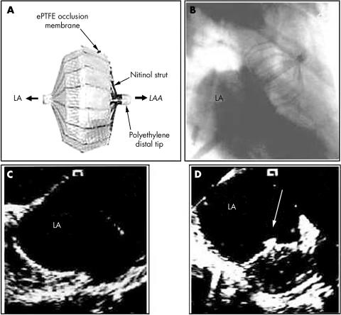
Percutaneous closure of the LAA. (A) Occluder device. (B) Fluoroscopic image of the occluder device in situ. (C) ICE image of the left atrium (LA) and LAA before deployment. (D) ICE following deployment of the device. Adapted and reproduced from Nakai et al, with permission.
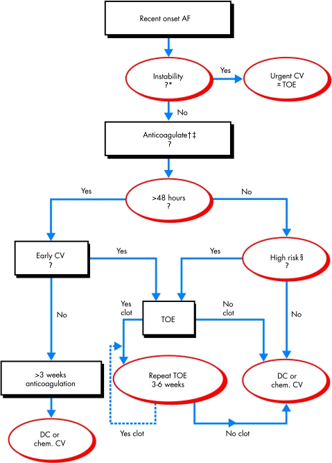
Proposed algorithm for the approach to patients presenting with recent onset atrial fibrillation. *Patients with haemodynamic instability, acute coronary syndrome, or severe heart failure. †If anticoagulation with heparin (unfractionated or low molecular weight) is contraindicated, consider transoesophageal echo guided cardioversion and review contraindication to anticoagulation if thrombus detected. ‡If not high risk and <48 hours, no anticoagulation is needed. §High risk if recent left atrial appendage thrombus, prior thromboembolism, > 65 years old, heart failure, hypertension, left ventricular dysfunction, significant valve disease, left atrial dilatation, increased risk of haemorrhagic complications, or suboptimal anticoagulation. AF, atrial fibrillation; DC, direct current; Early CV, indication or preference for early cardioversion (see text); TOE, transoesophageal echocardiogram.
References
-
- Peters NS, Schilling RJ, Kanagaratnam P, et al. Atrial fibrillation: strategies to control, combat, and cure. Lancet 2002;359:593–603. ▸ An excellent recent review of pathophysiology and current approaches to the management of atrial fibrillation. - PubMed
-
- Fuster V, Ryden LE, Asinger RW, et al. ACC/AHA/ESC guidelines for the management of patients with atrial fibrillation: executive summary. A report of the American College of Cardiology/American Heart Association task force on practice guidelines and the European Society of Cardiology committee for practice guidelines and policy conferences (committee to develop guidelines for the management of patients with atrial fibrillation) developed in collaboration with the North American Society of Pacing and Electrophysiology. Circulation 2001;104:2118–50. ▸ Recent policy guidelines for the management of AF produced after collaboration between the AHA, ACC, and ESC. - PubMed
-
- Kannel WB, Wolf PA, Benjamin EJ, et al. Prevalence, incidence, prognosis, and predisposing conditions for atrial fibrillation: population-based estimates. Am J Cardiol 1998;82:2N–9N. - PubMed
-
- Feinberg WM, Blackshear JL, Laupacis A, et al. Prevalence, age distribution, and gender of patients with atrial fibrillation. Analysis and implications. Arch Intern Med 1995;155:469–73. - PubMed
-
- Ommen SR, Odell JA, Stanton MS. Atrial arrhythmias after cardiothoracic surgery. N Engl J Med 1997;336:1429–34. - PubMed
Publication types
MeSH terms
LinkOut - more resources
Full Text Sources
Medical