Clinical Trial
. 2003 Nov;112(10):1506-20.
doi: 10.1172/JCI19301.

Abnormal germinal center reactions in systemic lupus erythematosus demonstrated by blockade of CD154-CD40 interactions

Affiliations
Clinical Trial

Abnormal germinal center reactions in systemic lupus erythematosus demonstrated by blockade of CD154-CD40 interactions

Amrie C Grammer et al. J Clin Invest. 2003 Nov.

Erratum in

  • J Clin Invest. 2007 Mar;117(3):835

Abstract

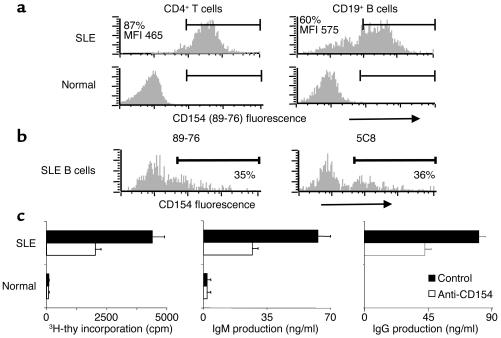
To determine the role of CD154-CD40 interactions in the B cell overactivity exhibited by patients with active systemic lupus erythematosus (SLE), CD19+ peripheral B cells were examined before and after treatment with humanized anti-CD154 mAb (BG9588, 5c8). Before treatment, SLE patients manifested activated B cells that expressed CD154, CD69, CD38, CD5, and CD27. Cells expressing CD38, CD5, or CD27 disappeared from the periphery during treatment with anti-CD154 mAb, and cells expressing CD69 and CD154 disappeared from the periphery during the post-treatment period. Before treatment, active-SLE patients had circulating CD38 (bright) Ig-secreting cells that were not found in normal individuals. Disappearance of this plasma cell subset during treatment was associated with decreases in anti-double-stranded DNA (anti-dsDNA) Ab levels, proteinuria, and SLE disease activity index. Consistent with this finding, peripheral B cells cultured in vitro spontaneously proliferated and secreted Ig in a manner that was inhibited by anti-CD154 mAb. Finally, the CD38(+/++)IgD(+), CD38(+++), and CD38(+)IgD(-) B cell subsets present in the peripheral blood also disappeared following treatment with humanized anti-CD154. Together, these results indicate that patients with active lupus nephritis exhibit abnormalities in the peripheral B cell compartment that are consistent with intensive germinal center activity, are driven via CD154-CD40 interactions, and may reflect or contribute to the propensity of these patients to produce autoantibodies.


Figures

Figure 1
Figure 1
Freshly isolated SLE B cells express functionally active CD154. (a and b) CD154 expression on CD4+ T cells and CD19+ B cells from the periphery of SLE patients and normal volunteers was assessed by flow cytometric staining of PBMCs for CD154 (PE-conjugated 89-76; shown in a and in b [left panel]; unconjugated 5c8 followed by PE-conjugated goat anti-mouse Ig, b [right panel]) and CD19 or CD4 (APC-conjugated mAb, a and b). The results of two of six experiments with similar findings are shown. (c) Freshly isolated, negatively selected peripheral B cells (0.1 × 10⁵) from active-SLE patients and normal volunteers were incubated in the presence of 10 μg/ml anti-CD154 (mIgG2a; 5c8) or 10 μg/ml control Ab (mouse IgG2a; P1.17). Analysis of DNA synthesis was carried out on day 3. Ig production was analyzed by ELISA after a 5-day incubation. All determinations were performed in duplicate and are expressed as the mean ± SEM. The results of one of three experiments with similar findings are shown. 3H-thy, 3H-thymidine.
Figure 2
Figure 2
CD38-expressing B cell subsets in active-SLE blood disappear following two treatments with humanized anti-CD154 (5c8). Freshly isolated mononuclear cells (MNCs) from active-SLE patients (a and b), normal volunteers (a), and individual tonsils (a) were assessed for CD19+ subpopulations by FACS analysis following staining with FITC-conjugated anti-IgD, PE-conjugated anti-CD38, and APC-conjugated anti-CD19. Freshly isolated PBMCs from active-SLE patients before treatment, during treatment (SLE nos. 1 and 2, 2 months; SLE nos. 3 and 4, 1 month), and after treatment withdrawal (SLE no. 1, 3 months after; SLE no. 2, 20 months after; SLE no. 3, 2 months after; SLE no. 4, 20 months after) were assessed for CD19+ subpopulations by FACS analysis following staining with FITC-conjugated anti-IgD, PE-conjugated anti-CD38, and APC-conjugated anti-CD19 (c). The mean ± SEM percentages of CD19+ B cells in each subset defined by CD38 and IgD are shown graphically. Statistical significance was determined by the Student’s t test and is depicted with each pair of symbols indicating a specific comparison: λP = 0.0445, αP = 0.0208, +P = 0.0208, βP = 0.00037, δP = 0.014, γP = 0.0197, *P = 0.016.
Figure 3
Figure 3
Expression of activation antigens by B cells diminishes with different kinetics following treatment of active-SLE patients with humanized anti-CD154 (5c8). Freshly isolated PBMCs from normal volunteers and active-SLE patients before treatment, during treatment, and after treatment (SLE no. 1, 2 months after; SLE no. 2, 20 months after; SLE no. 3, 3 months after; SLE no. 4, 20 months after) were assessed for CD69 and CD38 expression in CD19+ B cell subsets by FACS analysis following staining with FITC-conjugated anti-IgD, APC-conjugated anti-CD19, and PE-conjugated Ab against CD69 or CD38. The percentages of CD19+ pre-switch IgD+ and post-switch IgD B cells expressing CD38 (a) and CD69 (b) before, during, and after treatment are shown.
Figure 4
Figure 4
CD154 expression by active-SLE B cells after a treatment course with humanized anti-CD154 (5c8). Individual CD19+ B cells in freshly isolated PBMCs from normal volunteers and active-SLE patients before treatment, after two treatments, and 2–3 months after treatment were identified following staining with fluorochrome-conjugated anti-CD19 and either sorted into individual wells using a FACSVantage flow cytometer (a) or additionally stained with PE-conjugated anti-CD154 (89-76; b). Transcripts of CD154, CD40, and β-actin mRNA were identified by PCR and Southern blotting (a). Surface expression of CD154 is shown in b, where the dotted line indicates the cutoff for staining with a PE-conjugated isotype-matched control Ab.
Figure 5
Figure 5
Impact of treatment with humanized anti-CD154 Ab (5c8) on CD27-expressing B cell subsets. Freshly isolated PBMCs from active-SLE patients and normal volunteers were assessed for CD27 expression in B cell subsets by FACS analysis following staining with FITC-conjugated anti-CD27, PE-conjugated anti-IgD, and TC-conjugated anti-CD19. Histograms of CD27 expression on B cells from normal volunteers or active-SLE patients before treatment, during treatment (SLE nos. 1 and 2, 2 months; SLE no. 3, 1 month), or after withdrawal of treatment (SLE no. 1, 3 months after; SLE no. 2, 20 months after; SLE no. 3, 2 months after) are shown.
Figure 6
Figure 6
CD5 subsets of peripheral B cells diminish following treatment with humanized anti-CD154 Ab (h5c8). Freshly isolated PBMCs from active-SLE patients and normal volunteers were assessed for CD5 expression in B cell subsets by FACS analysis following staining with FITC-conjugated anti-IgD, PE-conjugated anti-CD19, and TC-conjugated anti-CD5. Histograms of CD5 expression on B cells from normal volunteers or active-SLE patients before treatment, during treatment (SLE nos. 1 and 2, 2 months; SLE no. 3, 1 month), or after withdrawal of treatment (SLE no. 1, 3 months after; SLE no. 2, 20 months after; SLE no. 3, 2 months after) are shown.
Figure 7
Figure 7
Anti-CD154 treatment eliminates proliferating B cells and Ig-secreting B cells from the periphery of active-SLE patients. (a) Freshly isolated PBMCs from active-SLE patients before treatment, after two treatments, and 2–3 months after the final treatment were assessed for cell cycle following permeabilization, fixation, and staining with propidium iodide and APC-conjugated anti-CD19 Ab. The percentages of live cells in the S, G2, or M stage are depicted. (b) Freshly isolated PBMCs from SLE no. 3 before treatment, after two treatments, and 3 months after the final treatment were assessed for intracellular Ig and cell cycle following permeabilization, fixation, and staining with propidium iodide and FITC-conjugated anti-Ig and APC-conjugated anti-CD19 Ab. The presence of IC Ig, the expression of CD38, and cell cycle status are depicted. The percentage of IC Ig+ B cells was determined by subtraction of the histogram generated for nonpermeabilized cells (surface Ig–positive) from the one generated for permeabilized cells (surface and IC Ig–positive) using CellQuest. The percentages of live cells that are CD38bright or IC Ig+ are depicted. SSC, side scatter; FSC, forward scatter.
Figure 8
Figure 8
Treatment of active-SLE patients with humanized anti-CD154 mAb (BG9588, 5c8) decreases anti-DNA Ab levels and the degree of proteinuria and improves SLEDAI. Changes in anti-dsDNA Ab levels, 24-hour urinary protein excretion (proteinuria), and SLEDAI are expressed on the left axis as percentage of base-line levels. Prednisone dose is shown on the right axis in mg/d. Vertical arrows indicate infusions of humanized anti-CD154 mAb (BG9588, 5c8). Horizontal arrows indicate additional immunosuppressive therapy. MMF, mycophenolate mofetil; CYC, cyclophosphamide.

Comment in

Similar articles

Cited by

References

    1. Grammer AC, Dorner T, Lipsky PE. Abnormalities in B cell activity and the immunoglobulin repertoire in human systemic lupus erythematosus. Molecular Pathology of Autoimmune Diseases. 2001;2:282–318.
    1. Lipsky PE. Systemic lupus erythematosus: an autoimmune disease of B cell hyperactivity. Nat. Immunol. 2001;2:764–766. - PubMed
    1. Grammer AC, Lipsky PE. CD154-CD40 interactions mediate differentiation to plasma cells in healthy individuals and persons with systemic lupus erythematosus. Arthritis Rheum. 2002;46:1417–1429. - PubMed
    1. Dorner T, Lipsky PE. Immunoglobulin variable-region gene usage in systemic autoimmune diseases. Arthritis Rheum. 2001;44:2715–2727. - PubMed
    1. Grammer AC, McFarland RD, Heaney J, Darnell BF, Lipsky PE. Expression, regulation, and function of B cell-expressed CD154 in germinal centers. J. Immunol. 1999;163:4150–4159. - PubMed

Publication types

MeSH terms