. 2003 Nov 25;100(24):14187-92.
doi: 10.1073/pnas.2332818100. Epub 2003 Nov 14.

Dietary Cu stabilizes brain superoxide dismutase 1 activity and reduces amyloid Abeta production in APP23 transgenic mice

Affiliations

Dietary Cu stabilizes brain superoxide dismutase 1 activity and reduces amyloid Abeta production in APP23 transgenic mice

Thomas A Bayer et al. Proc Natl Acad Sci U S A. .

Abstract

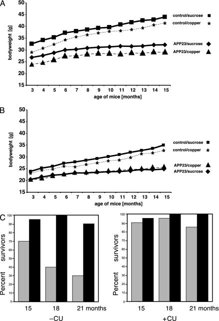
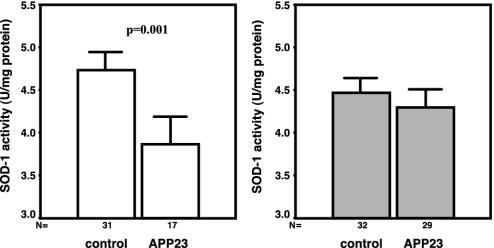
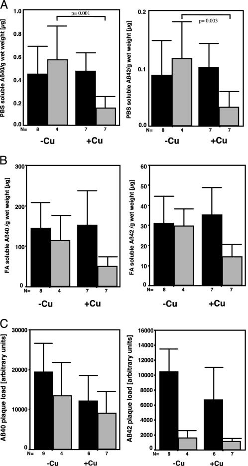
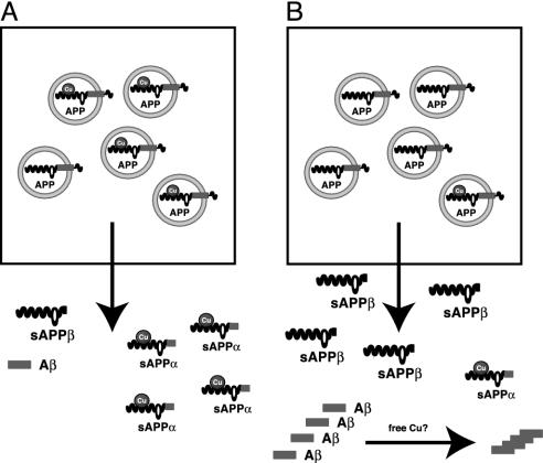
The Cu-binding beta-amyloid precursor protein (APP), and the amyloid Abeta peptide have been proposed to play a role in physiological metal regulation. There is accumulating evidence of an unbalanced Cu homeostasis with a causative or diagnostic link to Alzheimer's disease. Whereas elevated Cu levels are observed in APP knockout mice, APP overexpression results in reduced Cu in transgenic mouse brain. Moreover, Cu induces a decrease in Abeta levels in APP-transfected cells in vitro. To investigate the influence of bioavailable Cu, transgenic APP23 mice received an oral treatment with Cu-supplemented sucrose-sweetened drinking water (1). Chronic APP overexpression per se reduced superoxide dismutase 1 activity in transgenic mouse brain, which could be restored to normal levels after Cu treatment (2). A significant increase of brain Cu indicated its bioavailability on Cu treatment in APP23 mice, whereas Cu levels remained unaffected in littermate controls (3). Cu treatment lowered endogenous CNS Abeta before a detectable reduction of amyloid plaques. Thus, APP23 mice reveal APP-induced alterations linked to Cu homeostasis, which can be reversed by addition of dietary Cu.


Figures

Fig. 1.
Fig. 1.
Scheme of APP23 transgenic mice and nontransgenic littermate controls for the short-term treatment period of aged mice. APP23, APP transgenic mice; m, month.
Fig. 2.
Fig. 2.
Body weight analyses and survival rate of treated mice. Influence of 12 months of treatment on body weight of male (A) and female (B) mice. (C) Survival rate of APP23 and nontransgenic littermate control mice at 15, 18, and 21 months of age. All mice were treated for a 3-month period (short-term treatment) starting at 12, 15, and 18 months of age, respectively. APP23 mice showed a significant reduced life duration, which was not observed in the Cu-treated group. Cu had no detrimental effect on survival of nontransgenic control mice. APP23, APP transgenic mice (gray bars); wt, nontransgenic littermate controls (black bars).
Fig. 3.
Fig. 3.
Mean Cu levels and 95% confidence interval for mean (expressed as μg of Cu per g of wet weight) in brain homogenates of APP23 mice. Shown are Cu levels of aged APP23 mice at 15, 18, and 21 months of age, which were vehicle (–Cu)- or Cu (+ Cu)-treated for 3 months. A statistically significant difference is shown between vehicle- and Cu-treated APP23 mice (ANOVA).
Fig. 4.
Fig. 4.
Mean SOD-1 activity and 95% confidence interval for mean SOD-1 activity in brain homogenates of APP23 and nontransgenic littermate controls at 15, 18, and 21 months of age, which were vehicle (–Cu)- or Cu (+ Cu)-treated for 3 months. SOD-1 activity is expressed as mean activity (units/mg protein). A statistically significant difference was found between vehicle APP23 and wild-type mice with P = 0.001 as well as vehicle- and Cu-treated APP23 with P = 0.05 mice (ANOVA).
Fig. 5.
Fig. 5.
Mean values and 95% confidence interval for mean Aβ levels of Cu (+Cu)- or vehicle (–Cu)-treatment effects on PBS- and formic acid-soluble and plaque deposited Aβ40 and Aβ42 in APP23 mice at 15 months of age. (A) ELISA of PBS-soluble Aβ40 and Aβ42. (B) ELISA of formic acid-soluble Aβ40 and Aβ42. (C) The plaque load determined by densitometric measurement of aggregated Aβ40 and Aβ42 in senile plaques did not reveal differences between the treatment groups. The statistical difference between treatments for male mice is indicated (t test). For details see text. Open bars, female; filled bars, male.
Fig. 6.
Fig. 6.
Model for the metallobiology of APP. APP is involved in Cu homeostasis in brain with implications for AD. (A) Normal interaction of APP with Cu. Cu binds to the N-terminal domain of APP that is in the lumen of intracellular compartments. After fusion with the plasma membrane, Cu is released into the extracellular space as α-secretory APP–Cu complexes. In this case, APP-Cu is cleaved by the α-secretase close to the plasma membrane and then released. APP molecules without Cu are cleaved by β- and γ-secretase activities within intracellular compartments, which is probably due to a different conformation as compared with APP charged with Cu. Low levels of Aβ peptides are secreted from the cell. (B) Depletion of Cu, as observed in brains of AD patients and APP transgenic mice, enhances amyloidogenic processing of APP. Our data show reduced SOD-1 activity and Cu levels in brain tissue of APP transgenic mice that could both be rescued by Cu treatment. This is a coherent finding, because intracellular Cu availability recently turned out to be a critical determinant of SOD-1 activity (45). An enhanced release of Aβ peptides might act as a trap for Cu, although we did not find evidence for this notion in our in vivo studies.

References

    1. Selkoe, D. J. (2001) Physiol. Rev. 81, 741–766. - PubMed
    1. Tanzi, R. E., Gusella, J. F., Watkins, P. C., Bruns, G. A., St. George-Hyslop, P., Van Keuren, M. L., Patterson, D., Pagan, S., Kurnit, D. M. & Neve, R. L. (1987) Science 235, 880–884. - PubMed
    1. Kang, J., Lemaire, H. G., Unterbeck, A., Salbaum, J. M., Masters, C. L., Grzeschik, K. H., Multhaup, G., Beyreuther, K. & Muller-Hill, B. (1987) Nature 325, 733–736. - PubMed
    1. Dodart, J. C., Mathis, C. & Ungerer, A. (2000) Rev. Neurosci. 11, 75–93. - PubMed
    1. Hesse, L., Beher, D., Masters, C. L. & Multhaup, G. (1994) FEBS Lett. 349, 109–116. - PubMed

Publication types

MeSH terms