. 2004 Feb;15(2):543-51.
doi: 10.1091/mbc.e03-07-0487. Epub 2003 Nov 14.

Transcriptional activation of a constitutive heterochromatic domain of the human genome in response to heat shock

Affiliations

Transcriptional activation of a constitutive heterochromatic domain of the human genome in response to heat shock

Nicoletta Rizzi et al. Mol Biol Cell. 2004 Feb.

Abstract

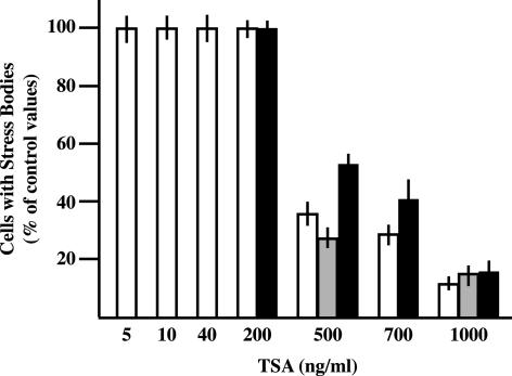
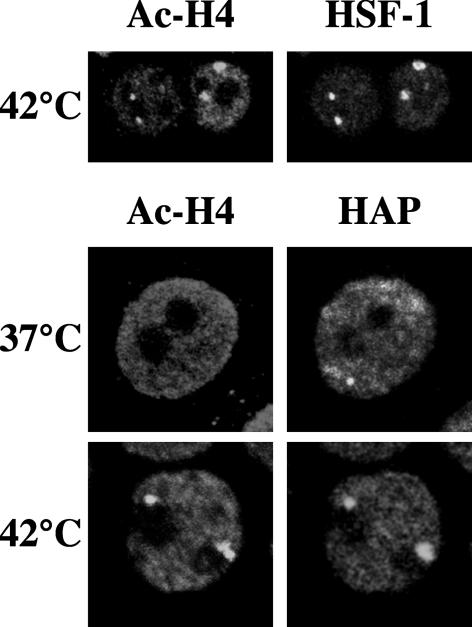
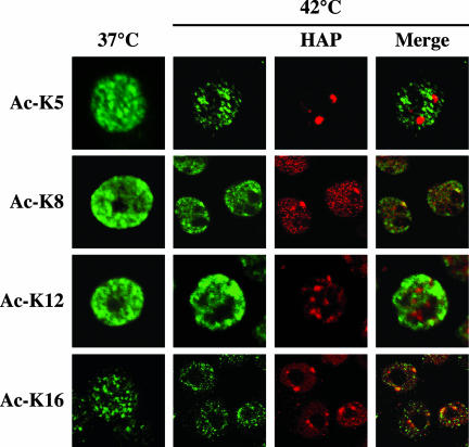
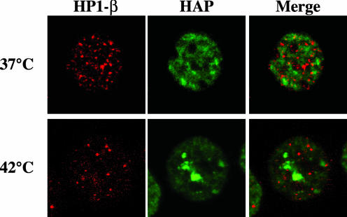
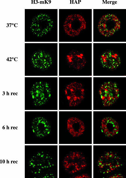
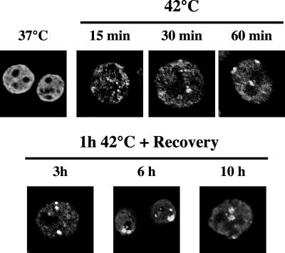
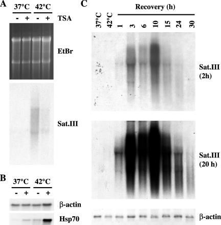
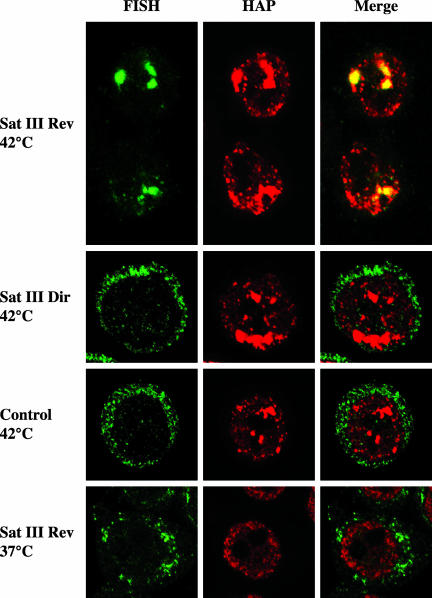
Heat shock triggers the assembly of nuclear stress bodies that contain heat shock factor 1 and a subset of RNA processing factors. These structures are formed on the pericentromeric heterochromatic regions of specific human chromosomes, among which chromosome 9. In this article we show that these heterochromatic domains are characterized by an epigenetic status typical of euchromatic regions. Similarly to transcriptionally competent portions of the genome, stress bodies are, in fact, enriched in acetylated histone H4. Acetylation peaks at 6 h of recovery from heat shock. Moreover, heterochromatin markers, such as HP1 and histone H3 methylated on lysine 9, are excluded from these nuclear districts. In addition, heat shock triggers the transient accumulation of RNA molecules, heterogeneous in size, containing the subclass of satellite III sequences found in the pericentromeric heterochromatin of chromosome 9. This is the first report of a transcriptional activation of a constitutive heterochromatic portion of the genome in response to stress stimuli.


Figures

Figure 1.
Figure 1.
TSA inhibits the assembly of stress bodies. HeLa cells were treated for 6 h with increasing concentrations of TSA. After removal of the drug, cells were heat-shocked 1 h at 42°C and then allowed to recover 1 h at 37°C before fixation with 4% formaldehyde. Cells were then independently stained with (1) rabbit polyclonal antibodies against hnRNP HAP, (2) mAb-96 against SF2/ASF, or (3) rat mAb 10H8 against HSF1. Primary antibodies were revealed with FITC-conjugated anti-rabbit, anti-mouse, or anti-rat goat antibodies. The fraction of cells with stress bodies was measured in three independent experiments counting 500 cells per experiments. This fraction was expressed as a percentage of the value measured in cells treated only with the solvent of TSA (ethanol). White bars, hnRNP HAP; gray bars, SF2/ASF; black bars, HSF1.
Figure 2.
Figure 2.
Stress bodies contain acetylated histone H4. HeLa cells were heat-shocked 1 h at 42°C and then allowed to recover 1 h at 37°C. Cells were then fixed in 4% formaldehyde and costained with a rabbit polyclonal antibody against the hyper-acetylated histone H4 and with either anti-HAP 16C3 mAb or the rat anti-HSF1 10H8 mAb. The distribution of hnRNP HAP and Ac-H4 in unstressed cells was also determined (37°C). Primary antibodies were revealed with FITC-conjugated anti-rabbit and with rhodamine-conjugated anti-rat or anti-mouse goat antibodies. Cells were then analyzed by confocal laser microscopy. Images of the same cells are shown.
Figure 3.
Figure 3.
Pattern of acetylation of histone H4 associated to stress bodies. Unstressed (37°C) or heat-shocked HeLa cells (1 h at 42°C followed by 1 h of recovery at 37°C) (42°C) were stained with rabbit polyclonal antibodies directed against different acetylated lysine residues of histone H4 as indicated. Primary antibodies were revealed with a FITC-conjugated anti-rabbit goat antibody. To reveal the localization of stress bodies, heat-shocked cells were costained with anti-HAP mAb 16C3 and with a rhodamine-conjugated anti-mouse goat antibody. Confocal laser microscopy images of representative cells are shown.
Figure 4.
Figure 4.
Stress bodies are not major sites of accumulation of HP1β. HeLa cells either unstressed (37°C) or heat-shocked 1 h at 42°C followed by 1 h at 37°C (42°C) were fixed in 4% formaldehyde. Cells were costained with a rabbit polyclonal antibody against hnRNP HAP, to reveal stress bodies, and with the rat mAb MAC353 specific for HP1β. Primary antibodies were revealed with FITC-conjugated anti-rabbit and rhodamine-conjugated anti-rat goat antibodies. Confocal laser microscopy images of the same fields are shown.
Figure 5.
Figure 5.
Stress bodies are not major sites of accumulation of histone H3 methylated on K9 (H3-mK9). HeLa cells either unstressed (37°C) heat-shocked 1 h at 42°C (42°C) or allowed to recover for the indicated time intervals were fixed in 4% formaldehyde. Cells were stained with the anti-HAP mAb 16C3 and with the rabbit antibody raised against the branched α-methH3-K9 peptide. Primary antibodies were revealed with FITC-conjugated anti-mouse and rhodamine-conjugated anti-rabbit goat antibodies. Confocal laser microscopy images of the same fields are shown.
Figure 6.
Figure 6.
Kinetics of histone H4 acetylation in stress bodies. HeLa cells were heat-shocked for 15, 30, or 60 min at 42°C. After 1 h of heat shock, cells were also allowed to recover at 37°C for the indicated time intervals. Cells were then stained with a rabbit polyclonal antibody against the hyper-acetylated form of histone H4 and with a FITC-conjugated anti-rabbit goat antibody. Confocal laser images of representative cells are shown. The distribution of histone H4 in unstressed HeLa cells is shown as a control (37°C).
Figure 7.
Figure 7.
Heat shock induces the production of satellite III transcripts. (A) HeLa cells were treated for 6 h with TSA (1 μg/ml; +) or with an equal volume of ethanol (-). After washing out the drug, cells were heatshocked 1 h at 42°C and allowed to recover 1 h at 37°C. Total RNAs were extracted and 10 μg were analyzed in Northern blotting with the pHuR98 probe specific for the subclass of satellite III sequences in the heterochromatic q1.2 band of human chromosome 9. The hybridization signal (overnight exposure) is shown along with an image of the gel stained with ethidium bromide to reveal the loaded material. (B) The same total RNAs described in A were hybridized to β-actin and hsp70 cDNA probes. (C) Total RNAs were prepared from unstressed cells (37°C) or from cells heat-shocked 1 h at 42°C (42°C) and then allowed to recover at 37°C for the indicated times. RNAs, 10 μg, were hybridized with the pHuR98 probe. Two exposures of the same filter are shown. As a control the filter was also hybridized to the β-actin cDNA.
Figure 8.
Figure 8.
Satellite III transcripts colocalize with hnRNP HAP in stress bodies. HeLa cells were heat-shocked 1 h at 42°C and then allowed to recover 3 h at 37°C. Cells were analyzed by in situ hybridization with oligonucleotides complementary to either the upper strand (Sat III Rev 42°C) or the lower strand (Sat III Dir 42°C) of the satellite III sequence. Heat-shocked cells were also hybridized to a control oligo complementary to a pBR322 sequence (Control 42°C). Finally the Sat III Rev 20-mer was hybridized to unstressed HeLa cells (Sat III Rev 37°C). The distribution of the hybridized oligos was revealed with fluorescein-avidin. The distribution of hnRNP HAP in the same cells was revealed with rabbit polyclonal anti-HAP and rhodamine-conjugated goat anti-rabbit antibodies. Confocal laser microscopy images of the same fields are shown.

References

    1. Agalioti, T., Chen, G., and Thanos, D. (2002). Deciphering the transcriptional histone acetylation code for a human gene. Cell 111, 381-392. - PubMed
    1. Boumil, R.M., and Lee, J.T. (2001). Forty years of decoding the silence in X-chromosome inactivation. Hum. Mol. Genet. 10, 2225-2232. - PubMed
    1. Brockdorff, N. (2002). X-chromosome inactivation: closing in on proteins that bind Xist RNA. Trends Genet 18, 352-358. - PubMed
    1. Chiodi, I., Biggiogera, M., Denegri, M., Corioni, M., Weighardt, F., Cobianchi, F., Riva, S., and Biamonti, G. (2000). Structure and dynamics of hnRNP-labelled nuclear bodies induced by stress treatments. J. Cell Sci. 113, 4043-4053. - PubMed
    1. Chirgwin, J.M., Przybyla, A.E., MacDonald, R.J., and Rutter, W.J. (1979). Isolation of biologically active ribonucleic acid from sources enriched in ribonuclease. Biochemistry 18, 5294-5299. - PubMed

Publication types

MeSH terms

LinkOut - more resources