. 2004 Feb;15(2):588-99.
doi: 10.1091/mbc.e03-07-0468. Epub 2003 Nov 14.

Gelsolin mediates collagen phagocytosis through a rac-dependent step

Affiliations

Gelsolin mediates collagen phagocytosis through a rac-dependent step

Pamela D Arora et al. Mol Biol Cell. 2004 Feb.

Abstract

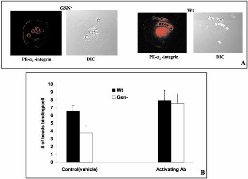
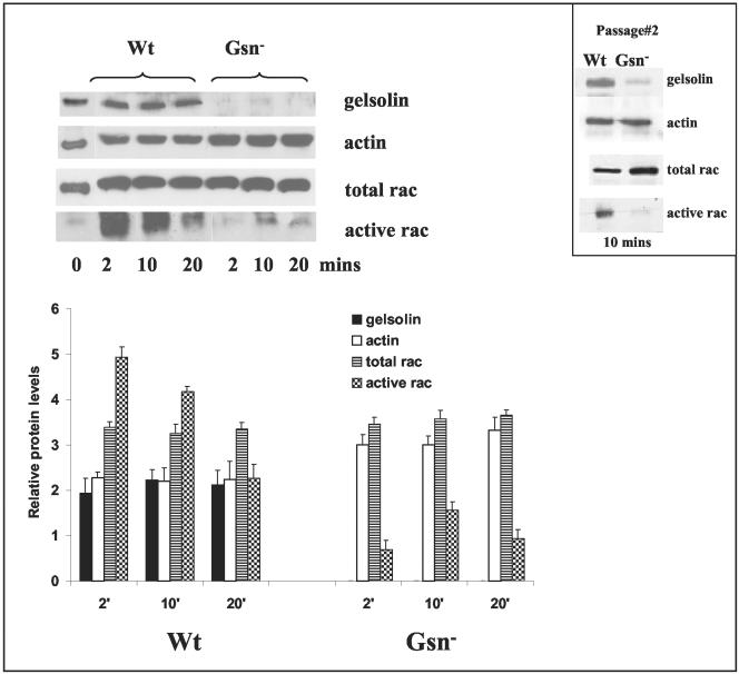
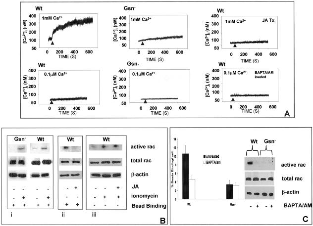
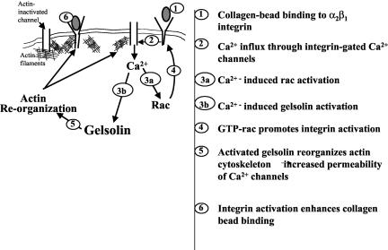
The role of gelsolin, a calcium-dependent actin-severing protein, in mediating collagen phagocytosis, is not defined. We examined alpha 2 beta 1 integrin-mediated phagocytosis in fibroblasts from wild-type (WT) and gelsolin knockout (Gsn(-)) mice. After initial contact with collagen beads, collagen binding and internalization were 60% lower in Gsn(-) than WT cells. This deficiency was restored by transfection with gelsolin or with beta1 integrin-activating antibodies. WT cells showed robust rac activation and increased [Ca(2+)](i) during early contact with collagen beads, but Gsn(-) cells showed very limited responses. Transfected gelsolin in Gsn(-) cells restored rac activation after collagen binding. Transfection of Gsn(-) cells with active rac increased collagen binding to WT levels. Chelation of intracellular calcium inhibited collagen binding and rac activation, whereas calcium ionophore induced rac activation in WT and Gsn(-) cells. We conclude that the ability of gelsolin to remodel actin filaments is important for collagen-induced calcium entry; calcium in turn is required for rac activation, which subsequently enhances collagen binding to unoccupied alpha 2 beta 1 integrins.


Figures

Figure 1.
Figure 1.
Involvement of gelsolin in the initial binding step of collagen phagocytosis. (A) Fibroblasts from WT mice were plated on collagen-coated beads for 20 min, fixed, and immunostained with polyclonal antibody to gelsolin. Cells show enrichment of gelsolin around collagen beads. DIC, differential interference contrast microscopy of same cells immunostained for gelsolin. (B) Gsn- and WT cells were plated at indicated collagen bead:cell ratios, incubated for 30 min, and collagen bead binding was analyzed by flow cytometry. There was significantly lower percentage of cells binding beads in Gsn- cells compared with WT cells. Data are mean ± SEs of mean percentage of cells binding collagen beads for B and C (n = 4 independent samples/data point). (C) Gsn- cells transfected with cDNA gelsolin expression vector for 48 h show gelsolin expression at ∼50% of WT cells. When analyzed with collagen bead binding assay, there was 50% enhancement of collagen bead binding in transfected cells. (D and E) α2β1-mediated collagen bead binding and phagocytosis in Gsn- and WT cells. Cells were plated on collagen-coated beads at 8:1 (bead:cell ratios) at 37°C for the indicated times, put on ice, and collagen bead attachment (D) and phagocytosis (E) were determined. Internalized beads were discriminated by quenching extracellular bound beads with trypan blue. Fluorescent and quenched beads were counted in 60 cells/sample at each time point. The numbers of beads binding per cell were significantly lower in Gsn- cells (p < 0.05), which also showed delayed bead internalization.
Figure 2.
Figure 2.
Involvement of actin filaments and actin binding proteins in collagen bead binding. (A) Incubation of cells with cytochalasin D reduces the percentage of cells binding collagen beads in both WT and Gsn- cells by >10-fold. Data are mean ± SEs of mean percentage of cells binding collagen beads (n = 4 independent samples/data point). (B) De novo actin assembly around beads. Cells were allowed to bind to collagen beads for 2, 10, and 20 min and subsequently treated with 0.2% OG-PHEM buffer for 2 s, incubated with rhodamine-actin monomers, fixed, and stained with FITC-phalloidin. There was reduced and much slower incorporation of actin monomers into actin filaments in Gsn- cells. Inset, WT cells incubated with collagen beads for 2 min show staining of actin filaments with FITC-phalloidin (left), rhodamine actin monomer incorporation into nascent filaments around beads (middle), and phase contrast showing collagen beads. (C) Accumulation of actin and actin binding proteins during early events of bead binding. Collagen-bead associated proteins were isolated at indicated time points and immunoblotted. From immunoblots normalized for bead counts and protein abundance (by Bio-Rad assay), there was marked reduction in amounts of actin, cortactin, and Arp3 associated with collagen beads in Gsn- cells. Data are mean ± SEs of blot density adjusted for number of beads in sample (n = 3/sample). Immunoblotting of whole cell lysates from unstimulated Gsn- and WT cells is shown for corresponding proteins at right side of panel.
Figure 3.
Figure 3.
Influence of gelsolin on integrin function. (A) Recruitment of α2-integrin (labeled with PE-conjugated antibody) around bound collagen beads was similar in Gsn- and WT cells, suggesting that integrin avidity was not affected by Gelsolin. (B) For assessment of antibody-induced integrin activation, Gsn- and WT cells were incubated with beads at 8:1 (bead:cell ratio) in the presence of β1-integrin activating antibody (9EG7) and analyzed by fluorescence microscopy. There was 50% enhancement in collagen bead binding in Gsn- cells but no change in WT cells (p < 0.01). Data are mean ± SEs of mean number of beads bound per cell.
Figure 4.
Figure 4.
Gelsolin impacts on rac activation. Gsn- and WT cell cultures at passage 4 showed equal expression levels of rac. For rac activation assay, cells were serum starved overnight and were stimulated with collagen-coated beads for the times indicated. Cell lysates were clarified by centrifugation and incubated with PAK-PBD-agarose beads for 1 h at 4°C. Beads were washed four times and analyzed on SDS-PAGE gels and probed with rac antibody. In spite of equal amounts of rac in the WT and Gsn- cells, rac activation was very low in Gsn- cells compared with WT. Histograms show mean ± SEs of blot density for indicated proteins after adjustment for total protein content (by Bio-Rad). Inset, results of similar experiment on cells from passage 2.
Figure 5.
Figure 5.
Rac activation overcomes the antagonizing effect of Gsn- on collagen bead binding. (A and B) Gsn- and WT cells were transfected with cDNA constructs for constitutively active rac (rac Q61) or for a dominant negative rac (rac N17) both of which express a c-myc tag. Cells were plated on collagen-coated beads for 1 h, washed, and fixed. Transfected cells were distinguished by immunostaining for c-myc. Rac activation enhances more than twofold collagen bead binding in Gsn- cells and increases collagen bead binding to levels similar to that of untransfected WT cells. The dominant negative rac construct reduces collagen bead binding to very low levels in both Gsn- and WT cells. (C) Immunoblots for indicated proteins using PAK binding assay described in Figure 4. Gsn- cells were transfected with gelsolin expression vector as indicated and incubated with collagen beads (or not). Note that only in cells transfected with gelsolin and incubated with collagen beads is there detectable activation of rac.
Figure 6.
Figure 6.
Role of calcium in rac activation. (A) Intracellular calcium transients were measured in single, fura 2/AM (3 μM)-loaded cells, detected by ratio fluorimetry at 346 nm/380 nm (excitation) and 510 nm (emission) in the presence (1 mM) or absence (0.1 μM) of CaCl2. Loaded cells were plated on collagen-coated beads and measurements were done after initial cell-bead binding and only in cells with attached beads. WT cells show robust calcium transients compared with Gsn- cells. To determine the importance of subcortical actin filaments on collagen-induced calcium increases, cells were incubated with JA (1 μM for 15 min) and during attachment to collagen beads, [Ca2+]i was measured by ratio fluorimetry. WT cells were also loaded with BAPTA/AM (3 μM in buffer containing 0.1 μM CaCl2) and measured by ratio fluorimetry. (B) Role of calcium in regulation of rac. i) Gsn- and WT cells were treated with ionomycin (2 μM; indicated by +) for 20 min, and rac activation was measured with PAK binding assay in the presence of collagen beads as described in Figure 4. ii) Effect of actin polymerization on activation of rac measured after bead binding in the JA- and vehicle-treated WT cells. iii) JA-treated WT cells were further treated with or without ionomycin showing effect on rac activation. (C) Collagen bead binding was measured in WT and Gsn- cells in low calcium buffer (0.1 μM CaCl2) with and without preincubation with BAPTA/AM as described above. Rac activation assays were performed as in B with or without BAPTA/AM preincubation.
Figure 7.
Figure 7.
Role of gelsolin in collagen phagocytosis. Gelsolin is required for remodeling of subcortical actin filaments that can regulate collagen-induced calcium entry. Calcium in turn regulates rac activation, which is required for enhanced binding of collagen by collagen receptors.

References

    1. Allen, L.A., and Aderem, A. (1996). Molecular definition of distinct cytoskeletal structures involved in complement- and Fc receptor-mediated phagocytosis in macrophages. J. Exp. Med. 184, 627-637. - PMC - PubMed
    1. Allen, P.G., and Janmey, P.A. (1994). Gelsolin displaces phalloidin from actin filaments. A new fluorescence method shows that both Ca2+ and Mg2+ affect the rate at which gelsolin severs F-actin. J. Biol. Chem. 269, 32916-32923. - PubMed
    1. Axelrod, D., Koppel, D.E., Schlessinger, J., Elson, E., and Webb, W.W. (1976). Mobility measurement by analysis of fluorescence photobleaching recovery kinetics. Biophys. J. 16, 1055-1069. - PMC - PubMed
    1. Arcaro, A. (1998). The small GTP-binding protein rac promotes the dissociation of gelsolin from actin filaments in the neutrophils. J. Biol. Chem. 273, 805-813. - PubMed
    1. Arora, P.D., Bibby, K.J., and McCulloch, C.A. (1994). Slow oscillations of free intracellular calcium ion concentration in human fibroblasts responding to mechanical stretch. J. Cell. Physiol. 161, 187-200. - PubMed

Publication types

LinkOut - more resources