. 2004 Feb;15(2):611-24.
doi: 10.1091/mbc.e03-05-0343. Epub 2003 Nov 14.

Rabip4' is an effector of rab5 and rab4 and regulates transport through early endosomes

Affiliations

Rabip4' is an effector of rab5 and rab4 and regulates transport through early endosomes

Michael A Fouraux et al. Mol Biol Cell. 2004 Feb.

Abstract

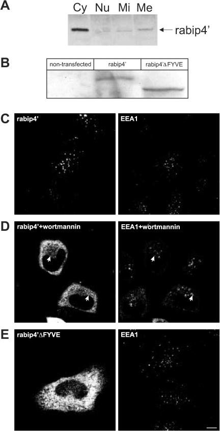
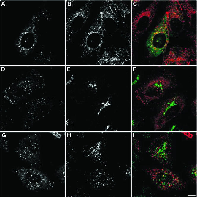
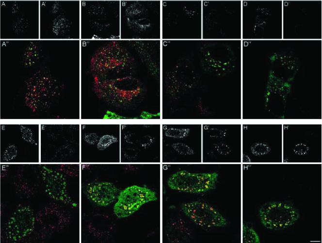
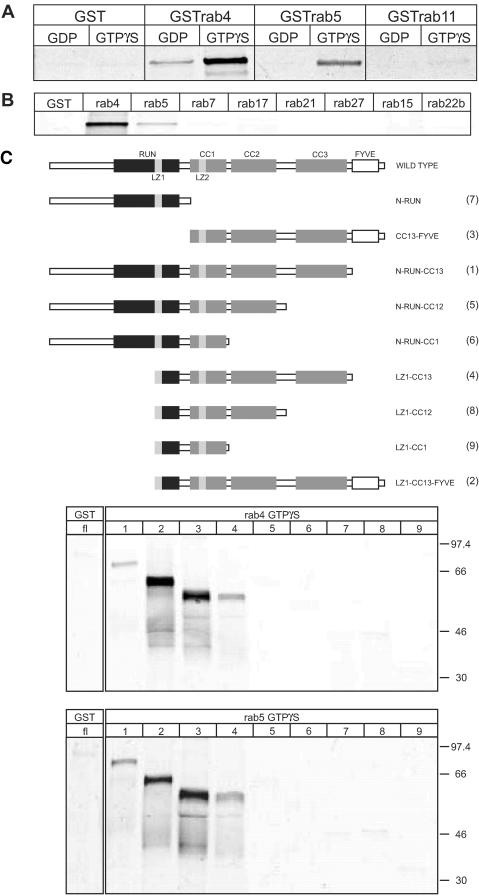
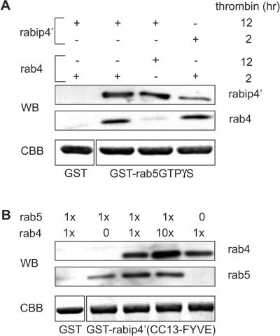
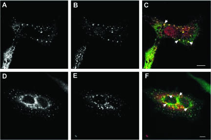
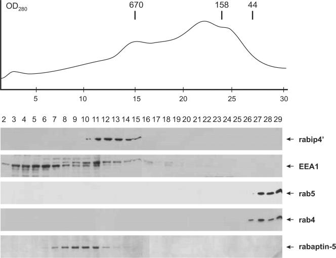
We describe the characterization of an 80-kDa protein cross-reacting with a monoclonal antibody against the human La autoantigen. The 80-kDa protein is a variant of rabip4 with an N-terminal extension of 108 amino acids and is expressed in the same cells. For this reason, we named it rabip4'. rabip4' is a peripheral membrane protein, which colocalized with internalized transferrin and EEA1 on early endosomes. Membrane association required the presence of the FYVE domain and was perturbed by the phosphatidylinositol 3-kinase inhibitor wortmannin. Expression of a dominant negative rabip4' mutant reduced internalization and recycling of transferrin from early endosomes, suggesting that it may be functionally linked to rab4 and rab5. In agreement with this, we found that rabip4' colocalized with the two GTPases on early endosomes and bound specifically and simultaneously to the GTP form of both rab4 and rab5. We conclude that rabip4' may coordinate the activities of rab4 and rab5, regulating membrane dynamics in the early endosomal system.


Figures

Figure 1.
Figure 1.
(A) Sequence and domain structure of rabip4′. Deduced amino acid sequence of rabip4′. Peptide sequences obtained by mass spectrometry are depicted in italics above the amino acid sequence. Leucine zipper motifs are underlined, the RUN motif is in italics, predicted coiled-coil domains are in bold, and the FYVE-finger domain is in bold italics. (B) Schematic structure of rabip4′. Coiled-coil domains are indicated with CC1, CC2, and CC3, the FYVE-finger domain with FYVE, leucine zippers with LZ1 and LZ2, and the RUN motif by RUN. (C) A Western blot of lysate from nontransfected HeLa cells (NT) was probed with an antibody against rabip4′(aa201-400) that detects 2 bands at 69 kDa and 80 kDa that were not seen by the preimmune (pi) serum. As positive controls, we used lysates of HeLa cells that were transfected with either VSV-G-rabip4′ or FLAG-RUFY1 and detected with epitope tag antibodies.
Figure 2.
Figure 2.
(A) The FYVE domain is required for membrane binding of rabip4′. HEp-2 cell lysates were fractionated as described in MATERIALS AND METHODS. Cytosolic (Cy), nuclear (Nu), mitochondrial (Mi), and microsomal (Me) fractions were analyzed by Western blotting by using a rabbit antibody against rabip4′. HeLa cells were transfected with VSV-G-tagged rabip4′ (B-D) or N-RUN-CC13 mutant (rabip4′ΔFYVE) lacking the FYVE domain (B and E). Cell lysates were resolved by SDS-PAGE and analyzed by Western blotting with an antibody against the epitope tag. Both constructs are expressed at the same level (B). HeLa cells expressing VSVG-tagged rabip4′ (C) were incubated for 15 min at 37°C with 50 nM wortmannin (D). Cells were fixed 18-24 h after transfection, doubly labeled with mouse anti VSV-G and rabbit anti-EEA1, stained with Alexa488-labeled rabbit anti-mouse and Cy3-labeled goat anti-rabbit, and examined by fluorescence microscopy. Note extensive colocalization between rabip4′ and EEA1 in C. In wortmannin-treated cells, rabip4′ redistributed partially in the cytoplasm but also colocalized with EEA1 on enlarged endosomes (arrows in D). Double-label immunofluorescence of cells transfected with VSV-G-tagged N-RUN-CC13 and endogenous EEA1 shows that the construct lacking the FYVE domain is localized in the cytoplasm (E). Bar, 10 μm.
Figure 3.
Figure 3.
rabip4′ is associated with early endosomes. HeLa cells were transiently transfected with VSV-G-tagged rabip4′. To label the TfR pathway, cells were incubated for 30 min at 37°C with 25 μg/ml Alexa594-Tf (A-C). The cells were fixed and subjected to confocal immunofluorescence microscopy. rabip4′ was labeled with a mAb against the VSV-G tag and counterstained with Alexa488-labeled goat anti-mouse IgG (A and C) or Cy3-labeled goat anti-mouse IgG (D, F, G, and I). TGN46 detection was with a sheep antibody and Alexa488-goat anti-sheep (E and F), whereas LAMP-1 was detected with a rabbit antibody against its cytoplasmic tail and stained with Alexa488-goat anti-rabbit IgG (H and I). Merged images are shown in C, F, and I. Bar, 10 μm.
Figure 4.
Figure 4.
rabip4′ regulates Tf transport through early endosomes. HeLa cells were transfected with VSV-G-tagged rabip4′ (A-D) or the VSV-G-tagged CC13-FYVE rabip4′ mutant (E-H). Cells were labeled with mouse anti-VSV-G tag followed by Alexa488-goat anti-mouse IgG (A-H). Alexa594-Tf was taken up for 30 min at 16°C, washed, and chased at 37°C for 0 min (A′ and E′), 10 min (B′ and F′), 20 min (C′ and G′), and 40 min (D′ and H′) in the presence of 50 mM Desferal. The corresponding merged images are shown in A″-H″. Bar, 10 μm.
Figure 5.
Figure 5.
rabip4′ FYVE domain does not affect endosome morphology and Tf recycling. Pulse-chase experiments in control HeLa cells (A and B) or HeLa cells transfected with HA-rabip4′-FYVE (C-D′) or GFP-FENS1-FYVE (E-F′). Cells expressing HA-rabip4′-FYVE were labeled with mouse anti-HA tag followed by Alexa488-goat anti-mouse IgG (C and D), whereas expression of GFP-FENS1-FYVE was visualized by the GFP signal (E and F). Alexa594-Tf was internalized for 30 min at 16°C, washed, and chased at 37°C for 0 min (A, C′, and E′), and 20 min (B, D′, and F′) in the presence of 50 mM Desferal. Arrows denote colocalization of Alexa594-Tf and GFP-FENS1-FYVE. Bar,10 μm.
Figure 6.
Figure 6.
rab4 and rab5 bind directly to rabip4′. (A) GST, GSTrab4, GSTrab5, and GSTrab11 were loaded with GTPγS or GDP and incubated overnight with35S-labeled VSV-G-tagged rabip4′. Beads were washed three times, and proteins were eluted with 25 mM reduced glutathione. Eluates were resolved on 10% SDS-PAA gels and analyzed by phosphorimaging. (B) GST, GSTrab4, GSTrab5, GSTrab7, GSTrab15, GSTrab17, GSTrab21, GSTrab22b, and GSTrab27 were loaded with GTPγS and incubated with 35S-labeled VSV-G-tagged rabip4′ as described in A. rabip4′ was eluted with glutathione and analyzed by SDS-PAGE and phosphorimaging. (C) Schematic representation of rabip4′ truncation mutants. See legend to Figure 1B for domain nomenclature. GSTrab4 and GSTrab5 were loaded with GTPγS. Binding and analysis of 35S-labeled VSV-G-tagged rabip4′ (fl) and truncations N-RUN-CC13 (1), LZ1-CC13-FYVE (2), CC13-FYVE (3), LZ1-CC13 (4), NRUN-CC12 (5), N-RUN-CC1 (6), N-RUN (7), LZ1-CC12 (8), and LZ1-CC1 (9) was done as described in MATERIALS AND METHODS.
Figure 7.
Figure 7.
rab4 and rab5 bind simultaneously to rabip4′. (A) GSTrab5 (loaded with GTPγS) and GST were immobilized on GSH beads and incubated with rabip4′ (CC13-FYVE). After washing, a second incubation was performed with rab4-GTPγS at a molar ratio rab5:rab4 of 5. Bound proteins were eluted, resolved by SDS-PAGE, and analyzed by Western blotting by using antibodies against rabip4′ (SN570) and rab4 (8091). Then, 0.5% of GSTrab5 and GST was loaded on a SDS-PAA gel and stained with Coomassie Brilliant Blue (CBB) to control for input in the assays. rab4 bound to the GSTrab5 beads in the presence rabip4′, but not to GST beads when these were incubated with rabip4′. Extending the thrombin cleavage time of GSTrab4 from 2 to 12 h inactivates GTPγS-loaded rab4 and reduced rab4-binding to background level. (B) GSTrabip4′(CC13-FYVE) and GST were immobilized on GSH beads and first incubated with rab5-GTPγS. After washing, increasing amounts of rab4-GTPγS (x stands for 50 μg) were added and beads were further incubated. Bound proteins were eluted, resolved by SDS-PAGE and analyzed by Western blotting with antibodies against rab4 (8091) and rab5 (4F11). CBB staining shows that similar amounts of GSTrabip4′ (CC13-FYVE) and GST have been used in each reaction (1% of the input). Note that addition of a 10-fold molar excess of rab4 does not compete out bound rab5.
Figure 8.
Figure 8.
rab4 and rab5 colocalize with rabip4′. HeLa cells expressing VSV-G-rabip4′ and rab4 (A-C), or VSV-G-rabip4′ and EGFP-rab5 (D-F) were labeled with a mAb against the VSV-G tag and counterstained with either Alexa488-goat anti-mouse IgG (A) or Alexa594-goat anti-mouse (D). rab4 was labeled with a rabbit antibody and stained with Cy3-goat anti-rabbit IgG (B), whereas rab5 was detected by EGFP fluorescence (E). Merged images are shown in C and F, and colocalization is indicated by arrows. Bar, 10 μm.
Figure 9.
Figure 9.
Cytosolic rabip4′ is associated with a large protein complex. A HeLa S100 extract was fractionated by Superdex 200 HR 10/30 gel filtration chromatography as described in MATERIALS AND METHODS. The position of marker proteins is indicated above the elution profile. Column fractions were analyzed by Western blotting with antibodies against rabip4′, EEA1, rab5, rab4, and rabaptin5. The positions of the various proteins are indicated with arrows.

Similar articles

Cited by

References

    1. Alber, T. (1992). Structure of the leucine zipper. Curr. Opin. Genet. Dev. 2, 205-210. - PubMed
    1. Bache, K.G., Brech, A., Mehlum, A., and Stenmark, H. (2003). Hrs regulates multivesicular body formation via ESCRT recruitment to endosomes. J. Cell Biol. 162, 435-442. - PMC - PubMed
    1. Bottger, G., Nagelkerken, B., and van der Sluijs, P. (1996). Rab4 and rab7 define distinct endocytic compartments. J. Biol. Chem. 271, 29191-29197. - PubMed
    1. Bucci, C., Parton, R., Mather, I., Stunnenberg, H., Simons, K., and Zerial, M. (1992). The small GTPase rab5 functions as a regulatory factor in the early endocytic pathway. Cell 70, 715-728. - PubMed
    1. Burd, C.G., and Emr, S.D. (1998). Phosphatidylinositol(3)-phosphate signaling mediated by specific binding to RING FYVE domains. Mol. Cell 2, 157-162. - PubMed

Publication types

MeSH terms

LinkOut - more resources