. 2004 Jan;15(1):345-58.
doi: 10.1091/mbc.e03-05-0334. Epub 2003 Nov 14.

Fusion between phagosomes, early and late endosomes: a role for actin in fusion between late, but not early endocytic organelles

Affiliations

Fusion between phagosomes, early and late endosomes: a role for actin in fusion between late, but not early endocytic organelles

Rune Kjeken et al. Mol Biol Cell. 2004 Jan.

Abstract

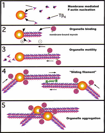
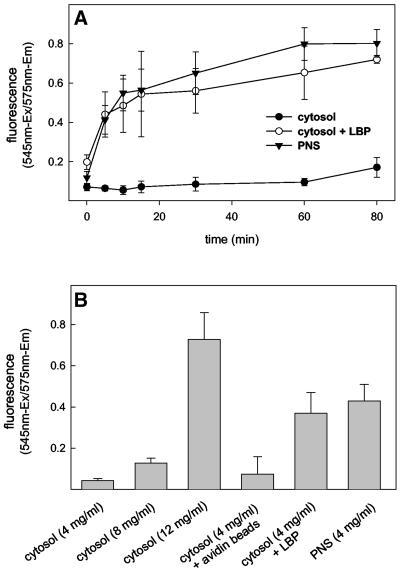
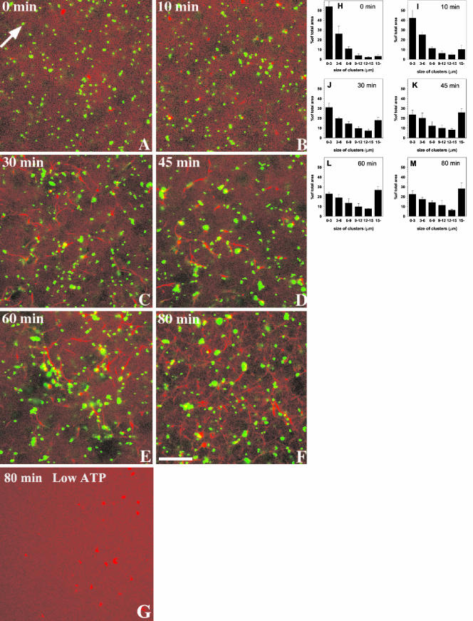
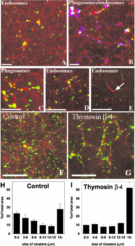
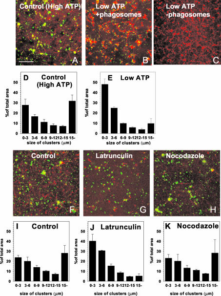
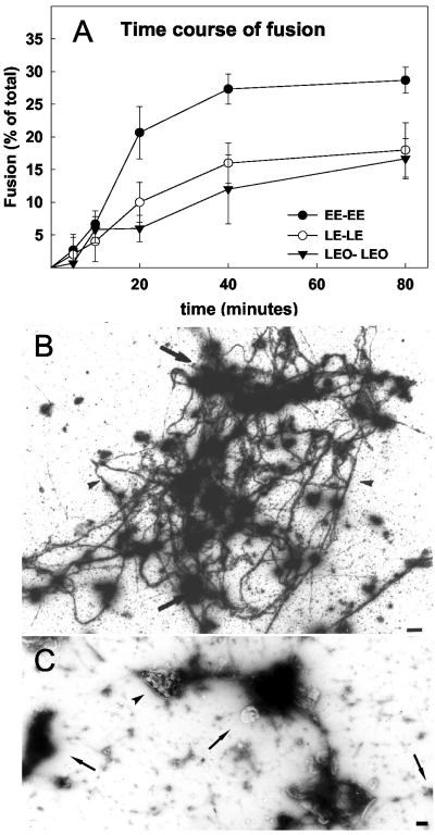
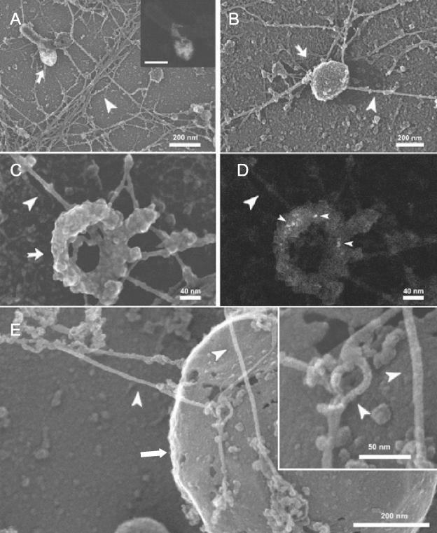
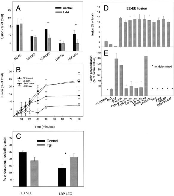
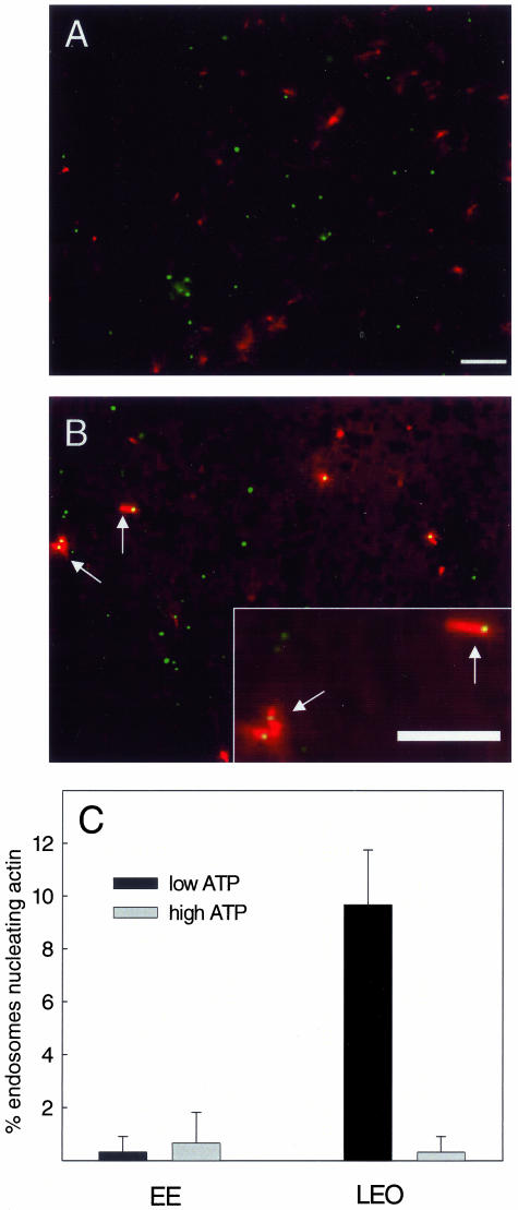
Actin is implicated in membrane fusion, but the precise mechanisms remain unclear. We showed earlier that membrane organelles catalyze the de novo assembly of F-actin that then facilitates the fusion between latex bead phagosomes and a mixture of early and late endocytic organelles. Here, we correlated the polymerization and organization of F-actin with phagosome and endocytic organelle fusion processes in vitro by using biochemistry and light and electron microscopy. When membrane organelles and cytosol were incubated at 37 degrees C with ATP, cytosolic actin polymerized rapidly and became organized into bundles and networks adjacent to membrane organelles. By 30-min incubation, a gel-like state was formed with little further polymerization of actin thereafter. Also during this time, the bulk of in vitro fusion events occurred between phagosomes/endocytic organelles. The fusion between latex bead phagosomes and late endocytic organelles, or between late endocytic organelles themselves was facilitated by actin, but we failed to detect any effect of perturbing F-actin polymerization on early endosome fusion. Consistent with this, late endosomes, like phagosomes, could nucleate F-actin, whereas early endosomes could not. We propose that actin assembled by phagosomes or late endocytic organelles can provide tracks for fusion-partner organelles to move vectorially toward them, via membrane-bound myosins, to facilitate fusion.


Figures

Figure 10.
Figure 10.
Hypothetical models to explain how action filaments nucleated from the surface of a membrane organelle could “attract” other myosin-bound organelles toward it. Because the barbed ends of actin are on, or close to the membrane surface, the polarity of the filaments is such that most myosins bound to the organelles would carry their cargo (small spheres) toward the nucleating organelle in the presence of ATP. In a second model, both fusion partners nucleate actin and the double-headed (bipolar) myosin II is proposed to cross-link and slide the filament bundles in opposite directions. This might also facilitate membrane organelle aggregation leading to docking and fusion.
Figure 1.
Figure 1.
F-Actin assembly during in vitro fusion conditions. (A) Kinetics of F-actin assembly. Macrophage cytosol alone (•) or with phagosomes (○) or PNS membranes (▾) were mixed and incubated in the presence of an ATP-regenerating system at 37°C. At the indicated time points, reaction was stopped and F-actin was quantified. (B) Summary of the total actin estimated under the given conditions after 80 min at 37°C. In A, each value represents the mean and range of two parallel tubes from one typical experiment. In B, each value represents the mean and SD of three parallel tubes from one typical experiment
Figure 2.
Figure 2.
Clustering of phagosomes along actin in macrophage cytosol in vitro. Phagosomes (green, false color) were mixed with freshly thawed macrophage cytosol mixed with rhodamine-G-actin (red) and incubated in the presence of an ATP-regenerating system at 37°C in a sealed chamber. The confocal microscopy images show a representative field after the indicated incubation times. The arrow in A indicates an individual phagosome. Structures larger than this are presumed to be clustered. Note the first signs of actin associated with LBP after 10-min incubation (arrow in B). (G) Macrophage cytosol mixed with rhodamine-G-actin (red) and incubated in the presence of an ATP-regenerating system at 37°C for 80 min in the absence of phagosomes. (H–M) Quantification of the data in A–F. The size of the clusters is indicated and the values are given as the percentage of the total area of green phagosomes. Bar, 10 μm.
Figure 3.
Figure 3.
Clustering of endocytic organelles and phagosomes along actin in macrophage cytosol and the role of Tβ4. PNS from cells pulsed for 40 min with Oregon Green-HRP (green) were mixed with macrophage cytosol containing rhodamine-G-actin plus ATP, and incubated at 37°C for 80 min (A). (B) Same experiment is shown with added phagosomes (shown in blue). (C and D) Higher magnifications of phagosomes (green) (C) and endocytic organelles (green) (D), aligning along actin bundles in the presence of ATP after 80-min incubation at 37°C. (E) Endocytic organelle (indicated by arrow) on the tip of an actin tail (comet). (G) Effects of 20 μM Tâ4 relative to untreated control (F). A quantification of the Tâ4 effect is shown in H and I. Bars, 10 μm.
Figure 4.
Figure 4.
Quantification of ATP- and actin-dependent phagosome clustering in macrophage cytosol and the role of microtubules. Phagosomes (green) were mixed with macrophage cytosol containing rhodamine-G-actin (red) and incubated for 80 min at 37°C with an ATP-regenerating system (A) or an ATP-depleting system (B). (C) Effect of an ATP-depleting system on F-actin in the absence of membranes. The clustering of phagosomes is quantified at high ATP levels (D) and low ATP levels (E). (F–H) Series of experiments with the same conditions as in A; control (F), 10 μM LatA (G), 10 μM nocodazole (H). The corresponding quantitative analyses are shown in I–K. All quantifications represent the mean values and SD of three separate experiments. Bars, 10 μm.
Figure 5.
Figure 5.
Kinetics of endocytic organelle fusion by on-grid EM and overview of structure. (A) Kinetics of this fusion at 37°C was determined by gold mixing by using EM. Mean and SD are shown from three separate experiments. (B) Low-magnification overview of the prominent actin cables (arrowheads) connected to many organelles (arrows), after 80-min incubation at 37°C with ATP. (C) Example of a fusion assay incubated in the presence of an ATP-depleting system for 80 min; gold-filled late endocytic organelles (arrows), actin (arrowheads). Bars, 1 μm.
Figure 6.
Figure 6.
On-grid EM fusion assay. (A) Principle of the EM fusion assay; 5-nm gold particles (small arrow) and 10-nm gold particles (small arrowhead) (derived from different sets of cells) are seen in the same (late) endocytic vesicle after 30-min pulse and 30-min chase in both fusion partners. The large arrowhead indicates a single actin filament oriented end-on to the organelle membrane, whereas the large arrow shows a bundle of actin to which the vesicle seems to be attached. (B–D) Examples of filaments putatively nucleated by late endocytic organelles. In B, three filaments (large arrowheads) are seen in a vesicle having only 5-nm gold particles (after 5-min incubation. (C) Two filaments (arrows) are seen in vesicle that has both small and large internalized gold particles (arrowheads) indicative of a fusion event after 10-min incubation. (D) More extensive filaments after 20-min incubation. This late vesicle contains two sizes of internalized gold (arrow) indicative of fusion, whereas the actin has been labeled with anti-actin and protein A gold (arrowhead). (E and F) Two examples of gold-filled late endocytic organelles (arrows) binding to F-actin bundles; the actin is labeled with anti-actin (arrowheads). Bars, 100 nm.
Figure 7.
Figure 7.
Cryo SEM of fusion assay. (A–D) Assay with endocytic organelles; A and B show an overview at low magnification; edocytic organelles is indicated by arrows; actin filaments are indicated by large arrowheads. The inset in A shows the back-scatter imaging mode to reveal the internalized gold. C shows details of actin filaments (arrowhead) impinging on an endocytic organelle (arrow); the corresponding bach-scatter image is seen in D; gold is indicated by small arrowheads. (E) Details of actin filaments (arrowheads) connected to a LBP (arrow) that is enlarged in the inset.
Figure 8.
Figure 8.
Quantification of fusion by EM and the biochemical EE fusion assay. (A) Comparison of the fusion between the different partner combinations indicated in the presence and absence of 10 μM LatA after 80-min incubation at 37°C by using Epon sections. (B) Kinetics of fusion of EE-EE and LEO-LEO with and without LatA (10 μM) by using the on-grid EM fusion assay. (C) Effect of 20 μM Tβ4 on the extent of fusion between LBP and either EE or LEO by EM by using Epon sections. Stars in A and C indicates significant differences p < 0.01 by using the Student's t test. (D) Effects of treatments that affect the F-actin on homotypic fusion between early endosomes in vitro using the biochemical assay. (E) Quantification of F-actin during some of the fusion reactions shown in Figure 9D. Each value in D and E represents the mean and SD of three parallel measurements from one typical experiment.
Figure 9.
Figure 9.
Nucleation of actin in the cytosol-free LM assay. Bodipyavidin–labeled EE (A) or LEO (B) were incubated with rhodamine actin and Tβ4 at low (0.2 mM) ATP. Actin is seen associated with a subset of LEO but not EE. Bar, 10 μm. (C) Quantitation of the percentage of EE and LEO that nucleate actin at low and high (5 mM) ATP.

Similar articles

Cited by

References

    1. Anes, E., Kuhnel, M.P., Bos, E., Pereira, J.M., Habermann, A., and Griffiths, G. (2003). Selected lipids activate phagosome actin assembly and maturation resulting in killing of pathogenic mycobacteria. Nat. Cell Biol. 5, 793-802. - PubMed
    1. Al-Haddad, A., et al. (2001). Myosin Va bound to phagosomes binds to F-actin and delays microtubule-dependent motility. Mol. Biol. Cell 12, 2742-2755. - PMC - PubMed
    1. Allen, L.H., and Aderem, A. (1995). A role for MARCKS, the alpha isozyme of protein kinase C and myosin I in zymosan phagocytosis by macrophages. J. Exp. Med. 182, 829-840. - PMC - PubMed
    1. Amann, K.J., and Pollard, T.D. (2000). Cellular regulation of actin network assembly. Curr. Biol. 10, R728-730. - PubMed
    1. Bader, M.F., Holz, R.W., Kumakura, K., and Vitale, N. (2002). Exocytosis: the chromaffin cell as a model system. Ann. N.Y. Acad. Sci. 971, 178-183. - PubMed

Publication types

LinkOut - more resources