. 2004 Feb;15(2):751-60.
doi: 10.1091/mbc.e03-05-0307. Epub 2003 Nov 14.

Mycobacterium tuberculosis phagosome maturation arrest: mycobacterial phosphatidylinositol analog phosphatidylinositol mannoside stimulates early endosomal fusion

Affiliations

Mycobacterium tuberculosis phagosome maturation arrest: mycobacterial phosphatidylinositol analog phosphatidylinositol mannoside stimulates early endosomal fusion

Isabelle Vergne et al. Mol Biol Cell. 2004 Feb.

Abstract

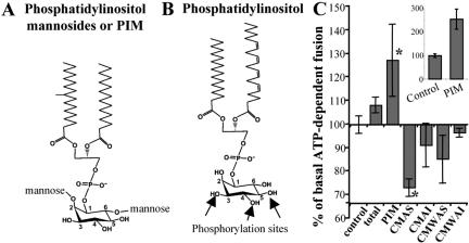
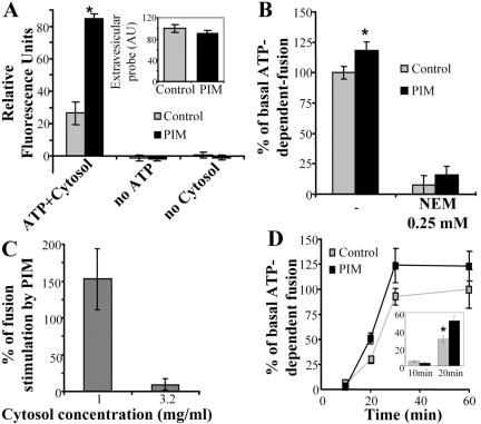
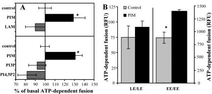
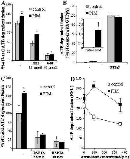
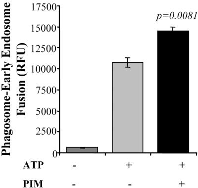
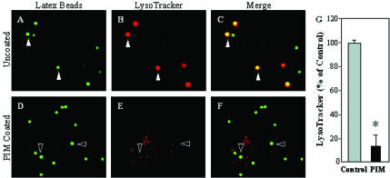
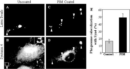
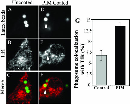
Mycobacterium tuberculosis is a facultative intracellular pathogen that parasitizes macrophages by modulating properties of the Mycobacterium-containing phagosome. Mycobacterial phagosomes do not fuse with late endosomal/lysosomal organelles but retain access to early endosomal contents by an unknown mechanism. We have previously reported that mycobacterial phosphatidylinositol analog lipoarabinomannan (LAM) blocks a trans-Golgi network-to-phagosome phosphatidylinositol 3-kinase-dependent pathway. In this work, we extend our investigations of the effects of mycobacterial phosphoinositides on host membrane trafficking. We present data demonstrating that phosphatidylinositol mannoside (PIM) specifically stimulated homotypic fusion of early endosomes in an ATP-, cytosol-, and N-ethylmaleimide sensitive factor-dependent manner. The fusion showed absolute requirement for small Rab GTPases, and the stimulatory effect of PIM increased upon partial depletion of membrane Rabs with RabGDI. We found that stimulation of early endosomal fusion by PIM was higher when phosphatidylinositol 3-kinase was inhibited by wortmannin. PIM also stimulated in vitro fusion between model phagosomes and early endosomes. Finally, PIM displayed in vivo effects in macrophages by increasing accumulation of plasma membrane-endosomal syntaxin 4 and transferrin receptor on PIM-coated latex bead phagosomes. In addition, inhibition of phagosomal acidification was detected with PIM-coated beads. The effects of PIM, along with the previously reported action of LAM, suggest that M. tuberculosis has evolved a two-prong strategy to modify its intracellular niche: its products block acquisition of late endosomal/lysosomal constituents, while facilitating fusion with early endosomal compartments.


Figures

Figure 1.
Figure 1.
Effects of mycobacterial lipids on early endosomal fusion. Structures of phosphatidylinositol mannosides PIM (A) and a typical mammalian phosphatidylinositol (B). PIM is glycosylated with single mannose residues at positions 2 and 6 of the inositol ring, whereas positions 3, 4, and 5 are unsubstituted. In eukaryotic cells, phosphatidylinositol can be phosphorylated at the positions 3, 4, or 5 on the inositol headgroup as indicated by arrows. (C) Early endosomal fusion was carried out in the presence of 40 μg/ml mycobacterial lipids, and 2 mg/ml cytosol, for 1 h at 37°C. CMAS and CMAI: lipids soluble in chloroform/methanol (2/1) (CM) and soluble (AS) or insoluble (AI) in acetone. CMWAS and CMWAI: lipids soluble in chloroform/methanol/water (10/10/3) (CMW) and soluble (AS) or insoluble (AI) in acetone. Total, unfractionated lipids extracted from M. tuberculosis. Inset, stimulatory effect of PIM upon preincubation of PIM with cytosol and ATP, before the initiation of endosomal fusion. Shown is the percentage of ATP-dependent fusion (1 mg/ml cytosol) in the presence or absence of PIM preincubated with cytosol and ATP-regenerating system for 20 min. Bars, SEM asterisk, p < 0.05 (ANOVA).
Figure 2.
Figure 2.
PIM stimulates early endosomal fusion in ATP-, NSF-, and cytosol-dependent manner. (A) In vitro fusion was carried out, in the presence or absence of 80 μg/ml PIM for 30 min. Reactions contained: 1 mg/ml cytosol or no cytosol (control); ATP regenerating system or an ATP-depleting system (no ATP). Inset, quantitation of extravesicular probes (avidin and biotinylated HRP), in the presence or absence of 80 μg/ml PIM. (B) NSF dependency of PIM-stimulated fusion. Endosomes and cytosol were preincubated for 20 min with 0.25 mM NEM followed by initiating fusion reaction with 1 mg/ml cytosol and incubation for 30 min. (C) Cytosol concentration effects on PIM-mediated stimulation of endosomal fusion. Fusion reaction was allowed to occur for 30 min in the presence of different concentrations of cytosol in the presence or absence of 80 μg/ml PIM. (D) PIM effects on rate and extent of endosomal fusion. Fusion was conducted in the presence of 2 mg/ml cytosol. Inset, percentage of ATP-dependent fusion in the presence or absence of PIM at 10 and 20 min. All percent values are given relative to the control fusion reaction without PIM. Bars, SEM asterisk, p < 0.05 (ANOVA).
Figure 3.
Figure 3.
Specificity of PIM-stimulated endosomal fusion. (A) Fusion was conducted in the presence of 40 μg/ml mycobacterial lipids (top) or 80 μg/ml lipids (bottom). (B) Fusion was conducted for 30 min, in the presence of 80 μg/ml PIM and 2 mg/ml cytosol, by using early endosomes (EE) or late endosomes (LE) loaded with biotinylated-HRP or avidin. Bars, SEM asterisk, p < 0.05 (ANOVA).
Figure 4.
Figure 4.
Analysis of the step at which PIM stimulates early endosomal fusion. Endosome preparations and cytosol, were preincubated for 20 min with indicated concentrations of GDI (A), GTPγS (25 μM) (B), BAPTA (C), or wortmannin (D). Fusion was carried out for 30 min, in the presence of 1 mg/ml cytosol (0.5 mg/ml cytosol was used for GTPγS experiment). Inset, percentage of ATP-dependent fusion (0.5 mg/ml cytosol) in the presence or absence of PIM. Bars, SEM asterisk, p < 0.05 (ANOVA).
Figure 5.
Figure 5.
PIM stimulates phagosome-early endosomes fusion in vitro. Phagosome, cytosol and endosome preparations were preincubated for 20 min at room temperature in absence or presence of 80 μg/ml PIM, and then fusion was carried out at 37C for 1 h in presence or absence of ATP. Bars, SEM.
Figure 6.
Figure 6.
PIM reduces acidification of latex bead phagosomes. Shown is epifluorescence microscopy of latex bead phagosomes in J774 cells. (A and D) Green fluorescence of latex beads. (B and E) LysoTracker staining carried out by incubating J774 cells with the dye 2 h before infection and throughout the experiment. (C and F) Merged images of green and red fluorescence. Yellow color illustrates the acidification of phagosomes. Cells contained either mock-treated control latex beads (A-C), or PIM-coated latex beads (D-F) as described in MATERIALS AND METHODS. Solid arrowheads exemplify colocalization of latex beads and LysoTracker staining. Hollow arrowheads exemplify phagosomes that are not acidified. (G) Quantitation of LysoTracker colocalization with phagosomes containing uncoated control beads or PIM-coated beads (30 min postinfection). The data are means ± SE of three separate experiments. *p < 0.001.
Figure 7.
Figure 7.
M. tuberculosis PIM causes syntaxin 4 accumulation on latex bead phagosomes. Shown is epifluorescence microscopy of latex bead phagosomes in J774 cells. (A and C) Green fluorescence of latex beads. (B and D) Red immunofluorescence of syntaxin 4. Latex beads were coated with purified M. tuberculosis H37Rv PIM and phagocytosed by J774 cells for 30 min. On phagocytosis, cells were fixed, permeabilized, and labeled with anti-syntaxin 4 antibody followed by Alexa 568-conjugated secondary antibody. Arrowheads indicated colocalization of latex beads with syntaxin 4. Quantitation of syntaxin 4 colocalization is shown numerically in (E). The data are means ± SE of three separate experiments (n = 826 phagosomes for control and n = 208 phagosomes for PIM).
Figure 8.
Figure 8.
M. tuberculosis PIM causes transferrin receptor accumulation on latex bead phagosomes. Shown is confocal microscopy of latex bead phagosomes in J774 cells. (A and D) Green fluorescence of latex beads. (B and E) Red immunofluorescence of transferrin receptor. (C and F) Merged images of green and red fluorescence. Latex beads were coated with purified M. tuberculosis H37Rv PIM and phagocytosed by J774 cells for 30 min. On phagocytosis, cells were fixed, permeabilized, and labeled with anti-transferrin receptor antibody followed by Alexa-568-conjugated secondary antibody. Arrowheads indicate colocalization of latex beads with transferrin receptor. Quantitation of transferrin receptor colocalization is shown numerically in G. The data are means ± SE of three separate experiments (n = 400 for control and n = 400 for PIM).

Similar articles

Cited by

References

    1. Anderson, R.J., and Roberts, E.G. (1930). The chemistry of lipoids of tubercule bacilli. J. Biol. Chem. 89, 611-611.
    1. Armstrong, J.A., and Hart, P.D.A. (1971). Response of cultured macrophages to M. tuberculosis with observations of fusion of lysosomes with phagosomes. J. Exp. Med. 134, 713-740. - PMC - PubMed
    1. Ballou, C.E., and Lee, Y.C. (1964). The structure of myoinositol mannoside from Mycobacterium tuberculosis. Biochemistry 72, 682-685. - PubMed
    1. Band, A.M., Ali, H., Vartiainen, M.K., Welti, S., Lappalainen, P., Olkkonen, V.M., and Kuismanen, E. (2002). Endogenous plasma membrane t-SNARE syntaxin 4 is present in rab11 positive endosomal membranes and associates with cortical actin cytoskeleton. FEBS Lett. 531, 513-519. - PubMed
    1. Barbieri, M.A., Hoffenberg, S., Roberts, R., Mukhopadhyay, A., Pomrehn, A., Dickey, B.F., and Stahl, P.D. (1998). Evidence for a symmetrical requirement for Rab5-GTP in in vitro endosome-endosome fusion. J. Biol. Chem. 273, 25850-25855. - PubMed

Publication types

MeSH terms

LinkOut - more resources