. 2003 Oct;203(4):379-89.
doi: 10.1046/j.1469-7580.2003.00222.x.

Effects of microinjected small GTPases on the actin cytoskeleton of human neutrophils

Affiliations

Effects of microinjected small GTPases on the actin cytoskeleton of human neutrophils

Margaret M Bird et al. J Anat. 2003 Oct.

Abstract

This paper describes a method for microinjection of proteins (Rho GTPases) into neutrophils and observations on the responses of the cells to these injections. Neutrophils are extremely difficult to inject because of their small size, complex morphology and fragility. To allow microinjections they must be cultured on a substrate that enables them to settle, adhere and spread. We determined that fibronectin- and/or collagen-coated coverslips are the best substrates and we used very fine needles and short microinjection times to minimize cell damage. These methods permitted us to inject up to 100 cells in a single preparation over a period of 30 min. Effects of microinjection were assessed by using tetramethylrhodamine isothiocyanate (TRITC)-phalloidin to label F-actin filaments, and observation by fluorescence and confocal scanning microscopy. Microinjection alone resulted in cell rounding and some changes in the F-actin cytoskeleton but injected cells remained adherent at the substrate, were able to respond to microinjected GTPases (V12Rac, V14RhoA, V12Cdc42) and continued to be responsive to activation by exposure to fMet-Leu-Phe (fMLP) or O-tetradecanoylphorbal 13-acetate (TPA). V12Rac caused an increase in neutrophil membrane ruffling and short protrusions from the cell membrane, whereas V14RhoA induced a large increase in punctate F-actin structures. V12Cdc42 produced focal condensation of F-actin and induced the formation of small microspikes. The differences between these responses of neutrophils and those of other similarly treated cell types are discussed. Our findings demonstrate that microinjection is a valuable technique for studying the role of individual proteins in neutrophils.


Figures
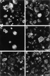
Fig. 1
Fig. 1
The influence of substrate on the settling and spreading of neutrophils on glass coverslips with and without additives, fixed and stained for F-actin after 60 min in culture. Adherence and spreading was only slightly better with poly-l-lysine (B) than with glass alone (A). Few cells settled on laminin (C) but those that did settle spread well. By contrast, cells settled and spread well on fibronectin (D) and collagen (E) separately or in a mixture of both (F). Scale bar = 10 µm.
Fig. 2
Fig. 2
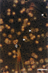
Phase contrast photograph showing neutrophils maintained on a collagen/fibronectin substrate with the microinjection needle (white arrow) in position ready to inject after 60 min in culture. All the flattened settled neutrophils within the field of view (black arrowheads) were injected. Scale bar = 20 µm.
Fig. 3
Fig. 3
Microinjected neutrophils retained the ability to respond to soluble stimuli such as fMLP and TPA. Cells were microinjected with rat IgG dissolved in microinjection buffer. Following microinjection, cells were stimulated with either 10 nm fMLP (A–C) or 100 ng mL−1 of TPA (D–F) for 5 min and then fixed with 3.7% formaldehyde in Ca2+/Mg2+-containing PBS. Microinjected cells were localized by the green fluorescence emitted after incubation with FITC-labelled anti-rat IgG and visualized as cells showing bright green fluorescence in the cytoplasm (A–Aii and D–Dii). F-actin localization was determined by labelling with TRITC-phalloidin (B–Bii and E–Eii) and visualized as red structures commonly located near the plasma membrane. Areas of co-localization of IgG and F-actin (C–Cii and F–Fii) are visualized as yellow structures. Arrowheads indicate the presence of podosomes; arrows indicate lamellipodia-like structures and asterisks indicate non-injected cells responding to respective stimuli. Control experiments in which injected cells were not exposed to either fMLP (Fig. 3A*–C*) or TPA (Fig. 3D*–F*) over an identical time span, showed that the cells remained rounded and lacked lamellipodia and podosomes. Scale bar = 10 µm.
Fig. 4
Fig. 4
The distribution of F-actin in neutrophils microinjected with different active and inactive forms of GTPases. Neutrophils were injected with IgG (A,B), or IgG plus V12Rac1 (C,D), V14RhoA (E,F), V12Cdc42 (G,H) and N17Rac1 (I,J). Injected cells were fixed in 3.7% formaldehyde 20 min after injections were completed. The actin cytoskeleton was visualized by staining with TRITC-labelled phalloidin (A,C,E,G,I) and microinjected cells were identified by FITC-labelled IgG (B,D,F,H,J). V12Rac injections caused some cell spreading, membrane ruffling and microspike formation; V14RhoA caused focal contact formation, cell spreading and microspike formation; and V12Cdc42 caused the most marked cell re-spreading, and some microspike formation. The inactive form, N17Rac1, caused some cell spreading but no microspike or lamellipodia formation. Scale bar = 10 µm.
Fig. 5
Fig. 5
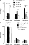
(A) Graph showing the responses of 100 neutrophils (fixed 20 min following the completion of the injections) to the microinjection of IgG, V12Rac1, V14RhoA and V12Cdc42. A significantly larger number of V12Cdc42-injected cells spread onto the substrate compared with both V12Rac1- (P < 0.0001) and V14RhoA- (P < 0.0001) injected cells. Microspike formation occurred in V12Rac1-, V14RhoA- and V12Cdc42-injected cells, but V14RhoA-injected cells had significantly larger numbers of microspikes than did Cdc42-injected cells (P < 0.0029). Lamellipodia formation occurred in significantly more V12Rac1-injected cells than both V14RhoA (P < 0.0001) and V12Cdc42 (P < 0.0001). V12Rho1-injected cells also formed significantly more lamellipodia than did V12Cdc42-injected cells (P < 0.0238). Control cells injected only with IgG remained significantly more rounded than both V12Rac1- and V12Cds42-injected cells, developed significantly fewer microspikes than V14RhoA-injected cells and significantly fewer lamellipodia than both V12Rac1- and V14RhoA-injected cells. *P < 0.05; ***P < 0.0001, †P < 0.005. (B) Graph showing the responses of 100 neutrophils (fixed for 20 min after the completion of the injection experiments) to the injection of IgG, N17Rac1, C3 transferase and N17Cdc42. A significantly larger number of cells injected with N17Rac1, C3 transferase and N17Cdc42 (P < 0.0001) spread onto the substrate compared with cells injected only with IgG. However, cells injected with N17Rac, C3 transferase and Cdc42 did not develop significantly larger numbers of either microspikes or lamellipodia.

References

    1. Allen WE, Jones GE, Pollard JW, Ridley AJ. Rho, Rac and Cdc42 regulate actin organization and cell adhesion in macrophages. J. Cell Sci. 1997;110:707–720. 10.1046/j.1469-7580.2003.00222.x. - DOI - PubMed
    1. Allen WE, Zicha D, Ridley AJ, Jones GE. A role for role for Cdc42 in macrophage chemotaxis. J. Cell Biol. 1998;141:1147–1157. 10.1046/j.1469-7580.2003.00222.x. - DOI - PMC - PubMed
    1. Aspenstrom P. The Rho GTPases have multiple effects on the actin cytoskeleton. Exp. Cell Res. 1999;246:20–25. 10.1046/j.1469-7580.2003.00222.x. - DOI - PubMed
    1. Bishop AL, Hall A. The Rho GTPases and their effector proteins. Biochem. J. 2000;348:241–255. 10.1046/j.1469-7580.2003.00222.x. - DOI - PMC - PubMed
    1. Burns S, Thrasher AJ, Blundell MP, Machesky L, Jones GE. Configuration of human dendritic cell cytoskeleton by Rho GTPases, the WAS protein, and differentiation. Blood. 2001;98:1142–1149. 10.1046/j.1469-7580.2003.00222.x. - DOI - PubMed

Publication types

Substances

LinkOut - more resources