

In situ hybridization and immunohistochemistry of versican, aggrecan and link protein, and histochemistry of hyaluronan in the developing mouse limb bud cartilage
- PMID: 14620382
- PMCID: PMC1571175
- DOI: 10.1046/j.1469-7580.2003.00226.x
In situ hybridization and immunohistochemistry of versican, aggrecan and link protein, and histochemistry of hyaluronan in the developing mouse limb bud cartilage
Abstract
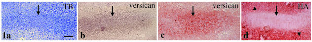
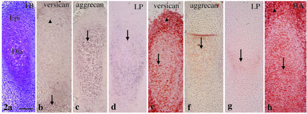
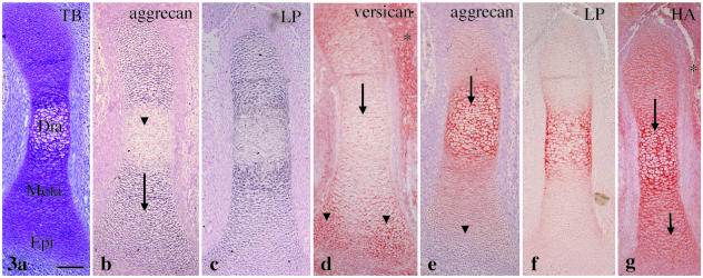
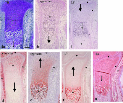
We investigated the expression pattern of versican, aggrecan, link protein and hyaluronan in the developing limb bud cartilage of the fetal mouse using in situ hybridization and/or immunohistochemistry. Versican mRNA and immunostaining were detected in the mesenchymal cell condensation of the future digital bone at E13. Versican mRNA expression rapidly disappeared from the tibial cartilage, as cartilage formation progressed during E13-15, but the immunostaining was gradually replaced by aggrecan immunostaining from the diaphysis. Immunostaining for both molecules thus had a 'nega-posi' pattern and consequently versican immunostaining was still detected at the epiphyseal end at E15. This result indicated that versican functions as a temporary framework in newly formed cartilage matrix. An aggrecan-positive region within the cartilage invariably had intense hyaluronan staining, whereas a versican-positive region also had affinity for hyaluronan within the cartilage, but not in the mesenchymal cell condensation. Therefore, the presence of versican aggregates was not confirmed in the developing limb bud cartilage. Furthermore, although link protein was more closely related with aggrecan than versican during limb bud cartilage formation, there was a discrepancy between the expression of aggrecan and link protein in tibial cartilage at E15. In particular, only a link protein-positive region was present in the marginal area of the metaphysis and the epiphysis at this stage. This finding may indicate a novel role for link protein.
Figures
Similar articles
-
Expression of versican in relation to chondrogenesis-related extracellular matrix components in canine mammary tumors.Histochem Cell Biol. 2005 Aug;124(2):139-49. doi: 10.1007/s00418-005-0007-y. Epub 2005 Sep 29. Histochem Cell Biol. 2005. PMID: 16088379
-
Histochemical localisation of versican, aggrecan and hyaluronan in the developing condylar cartilage of the fetal rat mandible.J Anat. 2001 Feb;198(Pt 2):129-35. doi: 10.1046/j.1469-7580.2001.19820129.x. J Anat. 2001. PMID: 11273038 Free PMC article.
-
Identification and characterization of versican/PG-M aggregates in cartilage.J Biol Chem. 2006 Jun 30;281(26):18257-63. doi: 10.1074/jbc.M510330200. Epub 2006 Apr 28. J Biol Chem. 2006. PMID: 16648631
-
[Cartilage proteoglycan aggregate: structure and function].Clin Calcium. 2004 Jul;14(7):9-14. Clin Calcium. 2004. PMID: 15577070 Review. Japanese.
-
Hyaluronic acid and hyaluronic acid-binding proteins in brain extracellular matrix.Anat Embryol (Berl). 1993 Nov;188(5):419-33. doi: 10.1007/BF00190136. Anat Embryol (Berl). 1993. PMID: 7508695 Review.
Cited by
-
Expression of versican in relation to chondrogenesis-related extracellular matrix components in canine mammary tumors.Histochem Cell Biol. 2005 Aug;124(2):139-49. doi: 10.1007/s00418-005-0007-y. Epub 2005 Sep 29. Histochem Cell Biol. 2005. PMID: 16088379
-
Hyaluronic acid is required for palatal shelf movement and its interaction with the tongue during palatal shelf elevation.Dev Biol. 2020 Jan 1;457(1):57-68. doi: 10.1016/j.ydbio.2019.09.004. Epub 2019 Sep 14. Dev Biol. 2020. PMID: 31526805 Free PMC article.
-
An in situ hybridization study of Runx2, Osterix, and Sox9 in the anlagen of mouse mandibular condylar cartilage in the early stages of embryogenesis.J Anat. 2008 Sep;213(3):274-83. doi: 10.1111/j.1469-7580.2008.00934.x. Epub 2008 Jul 8. J Anat. 2008. PMID: 18624832 Free PMC article.
-
Three-dimensional (3D) hydrogel serves as a platform to identify potential markers of chondrocyte dedifferentiation by combining RNA sequencing.Bioact Mater. 2021 Feb 23;6(9):2914-2926. doi: 10.1016/j.bioactmat.2021.02.018. eCollection 2021 Sep. Bioact Mater. 2021. PMID: 33718672 Free PMC article.
-
Morphogenetic and regulatory mechanisms during developmental chondrogenesis: new paradigms for cartilage tissue engineering.Tissue Eng Part B Rev. 2009 Mar;15(1):29-41. doi: 10.1089/ten.teb.2008.0329. Tissue Eng Part B Rev. 2009. PMID: 19063663 Free PMC article. Review.
References
-
- Asari A, Miyauchi S, Miyazaki K, Hamai A, Horie K, Takahashi T, et al. Intra- and extracellular localization of hyaluronic acid and proteoglycan constituents (chondroitin sulfate, keratan sulfate, and protein core) in articular cartilage of rabbit tibia. J. Histochem. Cytochem. 1992;40:1693–1703. 10.1046/j.1469-7580.2003.00226.x. - DOI - PubMed
-
- Bignami A, Perides G, Rahemtulla F. Versican, a hyaluronate-binding proteoglycan of embryonal precartilaginous mesenchyma, is mainly expressed postnatally in rat brain. J. Neurosci. Res. 1993;34:97–106. 10.1046/j.1469-7580.2003.00226.x. - DOI - PubMed
-
- Binette F, Cravens J, Kahoussi B, Haudenschild DR, Goetinck PF. Link protein is ubiquitously expressed in non-cartilaginous tissues where it enhances and stabilizes the interaction of proteoglycans with hyaluronic acid. J. Biol. Chem. 1994;269:19116–19122. 10.1046/j.1469-7580.2003.00226.x. - DOI - PubMed
-
- Bode-Lesniewska B, Dours-Zimmermann MT, Odermatt BF, Briner J, Heintz PU, Zimmermann DR. Distribution of the large aggregating proteoglycan versican in adult human tissues. J. Histochem. Cytochem. 1996;44:303–312. 10.1046/j.1469-7580.2003.00226.x. - DOI - PubMed
-
- Calabro A, Hascall VC, Caterson B. Monoclonal antibodies directed against epitopes within the core protein structure of the large aggregated proteoglycan (aggrecan) from the Swarm rat chondrosarcoma. Arch. Biochem. Biophys. 1992;298:349–360. 10.1046/j.1469-7580.2003.00226.x. - DOI - PubMed
Publication types
MeSH terms
Substances
Grants and funding
LinkOut - more resources
Full Text Sources
Molecular Biology Databases
