

Microtubule assembly in cultured myoblasts and myotubes following nocodazole induced microtubule depolymerisation
- PMID: 14620743
- PMCID: PMC1351055
Microtubule assembly in cultured myoblasts and myotubes following nocodazole induced microtubule depolymerisation
Abstract
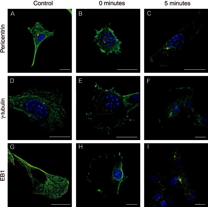
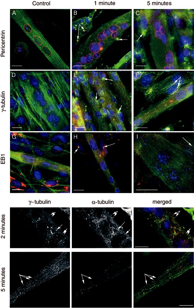
When myoblasts fuse into myotubes, the organisation of the cytoskeleton changes dramatically. For example, microtubules emanate in a radial array form the centrosome in myoblasts, but form linear arrays not linked to a centrosome in myotubes. It is not clear how these linear arrays are formed and nucleated. They could arise in a number of ways: by nucleation and release from centrosomal like structures, cytoplasmic assembly, breakage/severing or nucleation from non-centrosomal sites. To test which of the above mechanisms or combination of mechanisms are responsible we investigated the re-formation of microtubules after depolymerisation by nocodazole, using antibodies against pericentrin, gamma-tubulin, EB1, and tyrosinated alpha-tubulin. In myoblasts, we found that when microtubules were allowed to recover after complete depolymerisation with nocodazole, microtubule recovery began within 1 min and was complete after 5 min. Microtubules grew out from the centrosome, which was positively stained for gamma-tubulin or pericentrin. In untreated myotubes, microtubules were arranged in linear arrays, with EB1 at their ends. The pericentriolar protein, pericentrin was arranged in a band around the nucleus as well as discrete spots in the cytoplasm. In contrast, the microtubule nucleating protein gamma-tubulin was not found in a band around the nucleus, but was found in several punctuate spots throughout the cytoplasm. Further, when microtubules were allowed to recover, after complete depolymerisation with nocodazole, recovery was not as rapid as that seen in myoblasts, and we found that regrowth began with the formation of short microtubule fragments throughout the cytoplasm. Gamma-tubulin was associated with these fragments. These results suggest that in myotubes, nucleation of microtubules can be non-centrosomal.
Figures
Similar articles
-
Reorganization of microtubule nucleation during muscle differentiation.Cell Motil Cytoskeleton. 2005 Jan;60(1):1-13. doi: 10.1002/cm.20042. Cell Motil Cytoskeleton. 2005. PMID: 15532031
-
Control of microtubule nucleation and stability in Madin-Darby canine kidney cells: the occurrence of noncentrosomal, stable detyrosinated microtubules.J Cell Biol. 1987 Sep;105(3):1283-96. doi: 10.1083/jcb.105.3.1283. J Cell Biol. 1987. PMID: 2888771 Free PMC article.
-
Centrosomal deployment of gamma-tubulin and pericentrin: evidence for a microtubule-nucleating domain and a minus-end docking domain in certain mouse epithelial cells.Cell Motil Cytoskeleton. 1997;36(3):276-90. doi: 10.1002/(SICI)1097-0169(1997)36:3<276::AID-CM8>3.0.CO;2-5. Cell Motil Cytoskeleton. 1997. PMID: 9067623
-
Regulation of microtubule nucleation mediated by γ-tubulin complexes.Protoplasma. 2017 May;254(3):1187-1199. doi: 10.1007/s00709-016-1070-z. Epub 2017 Jan 10. Protoplasma. 2017. PMID: 28074286 Review.
-
Centrosomal and non-centrosomal microtubules.Biol Cell. 1999 May-Jun;91(4-5):321-9. Biol Cell. 1999. PMID: 10518998 Review.
Cited by
-
RacGAP50C directs perinuclear gamma-tubulin localization to organize the uniform microtubule array required for Drosophila myotube extension.Development. 2009 May;136(9):1411-21. doi: 10.1242/dev.031823. Epub 2009 Mar 18. Development. 2009. PMID: 19297411 Free PMC article.
-
Molecular robotic agents that survey molecular landscapes for information retrieval.Nat Commun. 2024 Apr 17;15(1):3293. doi: 10.1038/s41467-024-46978-2. Nat Commun. 2024. PMID: 38632239 Free PMC article.
-
Microtubule organization, dynamics and functions in differentiated cells.Development. 2017 Sep 1;144(17):3012-3021. doi: 10.1242/dev.153171. Development. 2017. PMID: 28851722 Free PMC article. Review.
-
Nuclear Mechanotransduction in Skeletal Muscle.Cells. 2021 Feb 4;10(2):318. doi: 10.3390/cells10020318. Cells. 2021. PMID: 33557157 Free PMC article. Review.
-
Cytoskeletal remodeling during myotube assembly and guidance: coordinating the actin and microtubule networks.Commun Integr Biol. 2009 Sep;2(5):452-7. doi: 10.4161/cib.2.5.9158. Commun Integr Biol. 2009. PMID: 19907716 Free PMC article.
References
Publication types
MeSH terms
Substances
Grants and funding
LinkOut - more resources
Full Text Sources