Comparative Study
. 2003 Oct;18(8):2273-84.
doi: 10.1046/j.l460-9568.2003.02973.x.

Networks of inhibitory and excitatory commissural interneurons mediating crossed reticulospinal actions

Affiliations
Comparative Study

Networks of inhibitory and excitatory commissural interneurons mediating crossed reticulospinal actions

B Anne Bannatyne et al. Eur J Neurosci. 2003 Oct.

Abstract

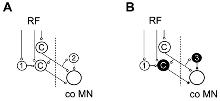
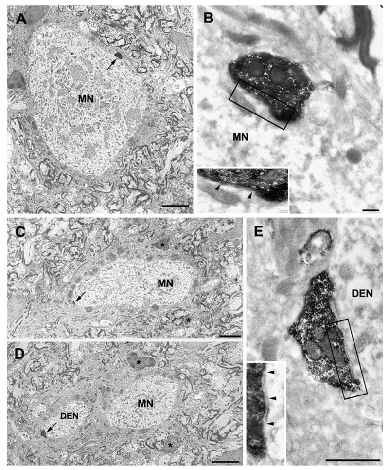
Axonal projections and neurotransmitters used by commissural interneurons mediating crossed actions of reticulospinal neurons were investigated in adult cats. Eighteen interneurons, located in or close to lamina VIII in midlumbar segments, that were monosynaptically excited by reticulospinal tract fibres and projected to contralateral motor nuclei were labelled by intracellular injection of tetramethylrhodamine-dextran and Neurobiotin. The nine most completely labelled interneurons were analysed with combined confocal and light microscopy. None of the stem axons gave off ipsilateral axon collaterals. Seven cells had axon collaterals that arborized in the contralateral grey matter in the ventral horn of the same segments. Transmitters were identified by using antibodies raised against vesicular glutamate transporters 1 and 2, glutamic acid decarboxylase and the glycine transporter 2. The axons of two cells were immunoreactive for the glycine transporter 2 and hence were glycinergic. Three cells were immunoreactive for the vesicular glutamate transporter 2 and hence were glutamatergic. None of the axons displayed immunoreactivity for glutamic acid decarboxylase. Electron microscopy of two cells revealed direct synaptic connections with motoneurons and other neurons. Axonal swellings of one neuron formed synapses with profiles in motor nuclei whereas those of the other formed synapses with other structures, including cell bodies in lamina VII. The results show that this population of commissural interneurons includes both excitatory and inhibitory cells that may excite or inhibit contralateral motoneurons directly. They may also influence the activity of motoneurons indirectly by acting through interneurons located outside motor nuclei in the contralateral grey matter but are unlikely to have direct actions on interneurons in the ipsilateral grey matter.


Figures

Fig. 1
Fig. 1
Possible connections in disynaptic and trisynaptic pathways between neurons in the reticular formation and contralateral motoneurons. Hypothetical circuits based on electrophysiological findings reported by Jankowska et al. 2003. (A) Connections in excitatory pathways. (B) Connections in inhibitory pathways. In both A and B, disynaptic connections are mediated via commissural interneurons (labelled C) that form synapses with contralateral motoneurons (co MN) whereas trisynaptic connections are either via ipsilateral interneurons (1) and the commissural interneurons represented by the lower neuron or via those represented by the upper commissural interneurons and other premotor interneurons including other commissural interneurons, contralateral excitatory interneurons (2) or contralateral inhibitory interneurons (3). Dotted vertical lines indicate the midline. White cells, excitatory; black cells, inhibitory.
Fig. 2
Fig. 2
Locations of cell bodies of RF commissural interneurons. (A and B) Locations of somata of interneurons located in the L4–L5 segments. Those used for confocal and EM analysis are indicated with filled circles.
Fig. 3
Fig. 3
Records and axonal projections of a glutamatergic interneuron. (A and B) Records from an interneuron (cell 4/1, Table 1) showing that it was monosynaptically activated from the reticular formation (RF; arrow indicates time of stimulus) and antidromically activated from the contralateral GS motor nucleus (co GS MN). Stimulation of group II afferents (from the quadriceps nerve stimulated at five times the threshold for fibres with the lowest threshold of activation: Q 5T) produced a polysynaptic IPSP. Averages of ten single records. In all of the cells illustrated, full spikes were generated only within the first few seconds after the penetration and the remaining blocked spikes usually consisted of an M spike or of M and IS spikes (of less than 10 mV). Upper traces show intracellular records and lower traces show records from the cord dorsum. Dotted lines in A indicate the first component of the descending volley from the reticular formation and the onset of the EPSP; note that latency of the EPSP with respect to the volley, i.e. the segmental latency, was <1ms as required for monosynaptic coupling. Dotted lines in B indicate the stimulus artefact and blocked antidromic spike potential evoked from the motor nucleus. Note that the latency was about 1 ms and too short for synaptically evoked responses. An additional record showing the RF volley at a higher amplification is shown below. Voltage calibrations (rectangular pulses at the beginning of records) and time calibrations (horizontal bars) are as indicated. (C) The location of the cell body and the course of its main axon (L, left; R, right side of cord). (D) Reconstruction of the axon collateral contained within the boxed area shown in C. Scale bar, 50 μm. The small boxed area (orientated by rotating clockwise by approximately 30°) contains the terminals examined with the confocal microscope shown in Fig. 4.
Fig. 4
Fig. 4
VGLUT2 immunoreactive axon terminals. (A) A projected confocal series of the collateral terminations (red) demarcated by the box in Fig. 3D. (B) A single optical section taken from the same series. (C) A single optical section showing immunoreactivity for VGLUT2 in the same plane as B (green). (D) A merged view of B and C. All axonal swellings viewed in this plane are immunoreactive for VGLUT2 but regions of preterminal axon do not contain the transporter. Arrows indicate individual boutons in A that are immunoreactive for VGLUT2 in the single optical section (C). Scale bar, 10 μm.
Fig. 5
Fig. 5
Records and axonal projections of a glycinergic interneuron. (A and B) Records from an interneuron (cell 1/5, Table 1) showing that a monosynaptic EPSP was evoked from the reticular formation (arrow indicates time of stimulus) and a blocked antidromic spike was evoked from the contralateral GS motor nucleus whereas stimulation of Q group II afferent fibres produced no response. Averages of ten single records. Format and abbreviations as in Fig. 3. Note that the latency of the monosynaptic EPSPs with respect to the first components of the descending volleys (< 1 ms) and the latency of the antidromic spike from the shock artefact (about 1 ms) were the same as for the cell illustrated in Fig. 3. Voltage calibrations (rectangular pulses at the beginning of records) and time calibrations (horizontal bars) are as indicated. (C) The location of the cell body and the course of its main axon (L, left; R, right side of cord). (D) Reconstruction of an axon collateral (boxed area in B). Scale bar, 50 μm. Terminals contained within the boxed area (1) were examined with the confocal microscope (Fig. 6).
Fig. 6
Fig. 6
GlyT2 immunoreactive axon terminals. (A) A confocal image showing a projected series of a collateral termination (red) contained within the boxed area shown in Fig. 5D. Arrows indicate boutons that appear in B–G. (B) A single optical section taken from the same series. Again the axon arbor is shown in red. (C) A further single optical section from the same plane showing immunoreactivity for GlyT2 (green). (D) A merged view of B and C. All axonal swellings in the single optical section are immunoreactive for GlyT2 (arrows), which typically is found on the axonal membrane surrounding boutons. Note that many of the axonal swellings arising from this collateral are clustered around the soma of a cell in lamina VII. (E–G) Single optical sections at higher magnification of boutons labelled 1–4 in C. Upper panels in each show interneuron boutons (red) and lower panels immunoreactivity for GlyT2 (green). Scale bars, 10 μm (A–D); 2.5 μm (E–G).
Fig. 7
Fig. 7
Records, axonal projections and immunoctyochemical characteristics of a glutamatergic interneuron also examined with the electron microsope. (A) Records of monosynaptic EPSPs evoked in an interneuron (cell 3/3, Table 1) from the reticular formation (arrows indicate times of stimuli) and of a blocked antidromic spike evoked from the contralateral GS motor nucleus. Both were followed by IPSPs. Averages of ten single records. Format and abbreviations as in Fig. 3. Note that the latency of the monosynaptic EPSPs with respect to the first components of the descending volleys (<1 ms) and the latency of the antidromic spike from the shock artefact (about 1 ms) were the same as for the cell illustrated in Fig. 3. Voltage calibrations (rectangular pulses at the beginning of records) and time calibrations (horizontal bars) are as indicated. (B) The location of the cell and the course of its main axon (L, left; R, right side of cord). (C) A reconstruction of the axon collateral (scale bar, 100 μm). The main axon gives rise to the collateral, which forms numerous terminations in lamina VII. Terminals contained within the boxed area (1) were examined with the confocal microscope. (D) The series of images shows single optical sections of two axonal swellings (red) and immunoreactivity for the vesicular glutamate transporter 2 (VGLUT2; green). The merged two-colour image shows that the terminals are VGLUT2-immunoreactive and hence that the cell is glutamatergic (scale bar, 10 μm). Terminals contained within the boxed area (2) were examined with the electron microscope (see Fig. 8).
Fig. 8
Fig. 8
Electron microscopy of glutamatergic terminals. (A) A light micrograph showing a cell contained within the boxed area 2 in Fig. 7C. A labelled collateral terminal is closely associated with it (arrow; scale bar, 10 μm). Two blood vessels are indicated (*) that are also present in the electron micrograph of the same area (B), which also shows the terminal (arrow; scale bar, 10 μm). C is a magnified view of the terminal (scale bar, 0.5 μm) and D shows details of the synaptic area (boxed area in C). The bouton forms an asymmetric synaptic arrangement with the cell (arrowheads).
Fig. 9
Fig. 9
Records and axonal projections of an interneuron examined with the electron microscope. (A) Records of monosynaptic EPSPs evoked in an interneuron (cell 3/5, Table 1) from the reticular formation (upper trace: arrows indicate times of stimuli), polysynaptic IPSP evoked from group II afferents and a blocked antidromic spike evoked from the contralateral GS motor nucleus (lower trace). Averages of ten single records. Note that the latency of EPSPs with respect to the first components of the descending volleys and of the spike potential with respect to the shock artefact was the same as for the cell illustrated in Fig. 3. Format and abbreviations as in Fig. 3. Voltage calibrations (rectangular pulses at the beginning of records) and time calibrations (horizontal bars) are as indicated. (B) The location of the cell and the course of its main axon (L, left, R, right side of cord). (C) Reconstruction of the axon collateral (boxed area in B). Note that this collateral ramifies principally in laminae VIII and IX. Axons within the boxed area (1) are illustrated in the electron micrographs shown in Fig. 10. Scale bar, 50 μm.
Fig. 10
Fig. 10
Electron micrographs of axon collaterals in lamina IX. (A) Low-power micrograph from the boxed area shown in Fig. 9C showing an axon terminal (arrow) adjacent to a motorneuron cell body (MN) in lamina IX. Scale bar, 10 μm. (B) A higher power image of the terminal with synaptic details (boxed area) shown in the inset (arrowheads). Scale bar, 0.5 μm. (C and D) Images taken further through serial sections of the same motorneuron (MN) around the point of origin of a large dendrite (DEN) showing a second labelled bouton associated with the cell (arrows; scale bars, 5 μm). The bouton formed a symmetrical synaptic contact (boxed area) with the dendrite (E, scale bar, 0.5 μm), details of which are shown in the inset (arrowheads). These synapses were studied in serial sections and sections were tilted to confirm that specializations were symmetrical.

Similar articles

Cited by

References

    1. Bannatyne BA, Maxwell DJ, Edgley SE, Hammar I, Jankowska E. Commissural interneurons in cat spinal motor pathways: identification of excitatory and inhibitory cells. J Physiol (Lond) 2003;5489:116.
    1. Bras H, Cavallari P, Jankowska E. Demonstration of initial axon collaterals of cells of origin of the ventral spinocerebellar tract in the cat. J Comp Neurol. 1988;273:584–592. - PubMed
    1. Bras H, Cavallari P, Jankowska E, Kubin L. Morphology of midlumbar interneurones relaying information from group II muscle afferents in the cat spinal cord. J Comp Neurol. 1989;290:1–15. - PubMed
    1. Cajal S, Ramon y. Histologie du Systeme Nerveux de L’homme et des Vertebres. Institute Cajal; Madrid: 1909.
    1. Drew T, Rossignol S. Functional organization within the medullary reticular formation of intact unanesthetized cat. I Movements evoked by microstimulation. J Neurophysiol. 1990a;64:767–781. - PubMed

Publication types

MeSH terms