. 2003 Nov 13;40(4):719-31.
doi: 10.1016/s0896-6273(03)00718-9.

Role of beta-catenin in synaptic vesicle localization and presynaptic assembly

Affiliations

Role of beta-catenin in synaptic vesicle localization and presynaptic assembly

Shernaz X Bamji et al. Neuron. .

Abstract

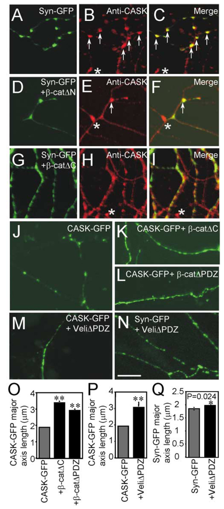
Cadherins and catenins are thought to promote adhesion between pre and postsynaptic elements in the brain. Here we show a role for beta-catenin in localizing the reserved pool of vesicles at presynaptic sites. Deletion of beta-catenin in hippocampal pyramidal neurons in vivo resulted in a reduction in the number of reserved pool vesicles per synapse and an impaired response to prolonged repetitive stimulation. This corresponded to a dispersion of vesicles along the axon in cultured neurons. Interestingly, these effects are not due to beta-catenin's involvement in cadherin-mediated adhesion or wnt signaling. Instead, beta-catenin modulates vesicle localization via its PDZ binding domain to recruit PDZ proteins such as Veli to cadherin at synapses. This study defines a specific role for cadherins and catenins in synapse organization beyond their roles in mediating cell adhesion.


Figures

Figure 1
Figure 1. Loss of β-Catenin in Hippocampal Pyramidal Neurons
(A) X-gal staining in the hippocampus of adult mice heterozygous for the CamKcre transgene and the R26R lacZ reporter. Counterstaining with Fast Red. (Left panels) (B, D, F, and H) 50 µm coronal sections from adult controls; (right panels) (C, E, G, and I) 50 µm coronal sections from adult β-catenin mutants. (B and C) β-catenin expression is lost from most (approximately 80%) CA3 pyramidal neurons in mutant mice. (D–G) β-catenin expression is absent in CA1 pyramidal neurons and the SR of mutant mice. (H and I) Synapsin expression is similar in the SR of control and mutant mice. Antibodies: rabbit polyclonal anti-β-catenin (B–E), mouse monoclonal anti-β-catenin (F and G), mouse monoclonal anti-synaptophysin (H and I). Scale bar, 450 µm (A); 35 µm (B–E); 6.5 µm (F–I).
Figure 2
Figure 2. Morphological and Functional Analyses of Hippocampal CA1 Synapses
(A–C) Electron microscope images of synapses in the SR of adult control (A) and β-catenin mutant (B and C) mice. (D–F) Synapses measured were those in which a welldefined postsynaptic density was observed (arrowheads). An area representing 700 µm2 of SR per animal was used for quantification of synapse density. The number of docked vesicles and undocked vesicles (vesicle cluster) was counted in at least 150 synapses per genotype. n = 4 per genotype. (G–L) Effects of β-catenin deletion on excitatory synaptic transmission in hippocampal CA1 synapses. Schaeffer collaterals were stimulated, and fEPSPs as well as fiber volleys were recorded in CA1 area of adult hippocampal slices. Open circles: wt; n = 5 animals. Filled triangles: MT; n = 4–6 animals. Data from multiple recordings of the same genotype were pooled and expressed as mean ± SEM. (G) Fiber volley amplitudes at different stimulation intensities. n = 13 for wt slices, and n = 10 for MT slices. (H) Input-output curves for basal synaptic transmission. The mean fEPSP slopes were plotted against fiber volley amplitude. The fiber volley amplitudes were binned, and corresponding EPSP slopes were averaged between slices. n = 13 for wt slices, and n = 10 for MT slices. (I) Paired-pulse facilitation (PPF) at low stimulation intensity (approximately 12.5% of maximal strength). Two stimuli were applied at various interpulse intervals (IPI, 10–100 ms). The ratios of the second and first EPSP slopes were calculated, and mean values are plotted against different IPIs. n = 9 for wt slices, and n = 8 for MT slices. (J) Posttetanic potentiation (PTP). A train of tetanic stimulation (100 Hz, 1 s, indicated by arrow) was applied to the CA1 synapses in the presence of the NMDA receptor antagonist Apv (100 µM), and EPSP slopes were plotted against time (4 min before and 10 min after the tetanus). n = 15 for wt slices, and n = 12 for MT slices. (K) Normal synaptic responses to a brief, high-frequency stimulation (HFS, 100 Hz, 40 pulses, in the presence of the NMDA receptor antagonist Apv, 100 µM) at CA1 synapses. n = 18 for wt slices, and n = 17 for MT slices. Inset: examples of EPSPs elicited by the HFS. (L) Reduced synaptic responses to prolonged repetitive stimulation (14 Hz, 80 pulses, Apv, 100 µM) in MT (n = 21 slices) compared to wt (n = 18 slices). Inset: superimposed EPSPs at 20th and 80th pulses. Asterisks denote significant differences from controls (p < 0.05). wt, wild-type; MT, β-catenin mutant mice. Scale bar, 250 nm (A and B); 190 nm (C).
Figure 3
Figure 3. Presynaptic Assembly Is Affected in Cultured Hippocampal Neurons Lacking β-Catenin
(A–C) Confocal images of rat hippocampal neurons transfected with synaptophysin-GFP (A) show colocalization with synaptotagmin ([B and C], arrows). (D–R) Confocal images of hippocampal neurons cultured from homozygous β-catenin flox mice. (D–F) Neurons transfected with synaptophysin-GFP (D) and immunostained with anti-Bassoon (E) show colocalization of synaptophysin-GFP and Bassoon puncta ([E and F], arrows). (G–O) Neurons expressing cre exhibit a diffuse pattern of synaptophysin-GFP expression (G, J, and M). Endogenous synaptotagmin is also diffusely expressed in neurons lacking β-catenin (H and I), whereas Bassoon ([K and L], arrows) and N-cadherin ([N and O], arrows) expression remains punctate. (P–R) PSD-95-GFP expression is normal and colocalizes with synaptophysin in neurons lacking β-catenin. Asterisks indicate immunopositive puncta on cells not transfected with synaptophysin-GFP or PSD-95-GFP. (S) Diffusion of synaptic vesicles was quantified by measuring the average length of the major axis of synaptophysin-GFP fluorescence in cells expressing synaptophysin-GFP alone or coexpressing the cre recombinase. (T) The density of Bassoon puncta in cells expressing synaptophysin-GFP or synaptophysin-GFP plus cre was quantified and expressed as the number of puncta per 10 µm neurite length ± SE. (U) The size of PSD-95-GFP fluorescence was quantified in cells expressing PSD-95-GFP alone or coexpressing the cre recombinase. Student’s t tests were performed, and asterisks indicate p values of <0.05. Scale bar, 10 µm.
Figure 4
Figure 4. Localization of Synaptophysin-GFP Fluorescence Is More Dynamic over Time in Neurons Lacking β-Catenin
Time-lapse confocal images of synaptophysin-GFP in β-catenin flox cultures in the absence of cre (A) or in cells expressing cre (B). Arrows mark dramatic changes in synaptophysin-GFP localization over time. Scale bar, 5 µm.
Figure 5
Figure 5. The PDZ Binding Domain of β-Catenin Is Important for Synaptic Vesicle Localization
(A) Protein binding regions and distinct domains of β-catenin. (B) The average length of the major axis of synaptophysin-GFP fluorescence was measured in cells expressing synaptophysin-GFP alone or coexpressing synaptophysin-GFP plus the indicated β-catenin. (C–L) Confocal images of rat hippocampal cultures. A punctate pattern of synaptophysin-GFP expression is observed in cells expressing synaptophysin-GFP alone (C) or coexpressing g-catAN (D). Synaptophysin-GFP expression is more diffuse in cells expressing β-catΔARM (E), β-catΔPDZ (F), and β-catΔAC (I). (F–H) Expression of β-catΔPDZ results in diffuse immunostaining of synaptotagmin that coincides with that of synaptophysin-GFP. (IΔK) Although expression of β-catΔC results in a diffuse pattern of synaptophysin-GFP staining, the pattern of bassoon immunostaining remains punctate. (L and M) Neurons expressing GFP plus either β-catΔC (L) or β-catΔPDZ (M) and immunostained with anti-synaptophysin. Note the punctate localization of synaptophysin in wild-type cells making contacts with neurons in which β-catenin function is inhibited. Asterisks indicate p values of <0.05 calculated using Student’s t tests (compared to synaptophysin-GFP alone). Scale bar, 10 µm (C–K); 3 µm (L and M).
Figure 6
Figure 6. Rescue of Synaptic Vesicle Phenotype in Neurons Lacking β-Catenin
(A–E) Confocal images of hippocampal neurons cultured from homozygous β-catenin flox mice, transfected with synaptophysin-GFP, cre, and mutant β-catenin. Expression of full-length β-catenin and β-catΔN restores the punctate pattern of synaptophysin-GFP staining (C and D), reflected by the decreased average length of synaptophysin-GFP fluorescence along the axon compared to cells expressing cre alone (F). Expression of β-catΔPDZ does not affect synaptophysin-GFP expression following β-catenin excision (E), and the average length of synaptophysin-GFP fluorescence is similar to that in cells expressing cre alone (F). Scale bar, 10 µm.
Figure 7
Figure 7. PDZ Protein Function in Synaptic Localization of Vesicles
(A–N) Confocal images of rat hippocampal cultures. (A–I) Neurons transfected with synaptophysin-GFP alone (A) or cotransfected with β-catΔN (D) or β-catΔC (G) were immunostained with anti-CASK (B, E, and H). Synaptophysin-GFP and CASK were colocalized in cells expressing synaptophysin-GFP alone ([C], arrows) or coexpressing β-catΔN ([F], arrows). CASK immunoreactivity was more diffusely expressed in cells expressing β-catΔC (H and I). Asterisks indicate CASK-positive puncta on cells not transfected with synaptophysin-GFP. CASK-GFP expression is punctate in cells expressing CASK-GFP alone (J) but is more diffuse in cells coexpressing either β-catΔC (K) or β-catΔPDZ (L). Expression of VelitΔPDZ results in a dramatic diffusion of CASK-GFP (M) but not of synaptophysin-GFP (N).(O–Q) Diffusion of CASK-GFP or synaptophysin-GFP was quantified by measuring the average length of the major axis of fluorescent puncta. Student’s t tests were performed, and asterisks indicate a p value of <0.05. Scale bar, 10 µm.

References

    1. Ahmari SE, Buchanan J, Smith SJ. Assembly of presynaptic active zones from cytoplasmic transport packets. Nat. Neurosci. 2000;3:445–451. - PubMed
    1. Barth AI, Nathke IS, Nelson WJ. Cadherins, catenins and APC protein: interplay between cytoskeletal complexes and signaling pathways. Curr. Opin. Cell Biol. 1997;9:683–690. - PubMed
    1. Benson DL, Tanaka H. N-cadherin redistribution during synaptogenesis in hippocampal neurons. J. Neurosci. 1998;18:6892–6904. - PMC - PubMed
    1. Bozdagi O, Shan W, Tanaka H, Benson DL, Huntley GW. Increasing numbers of synaptic puncta during late-phase LTP: N-cadherin is synthesized, recruited to synaptic sites, and required for potentiation. Neuron. 2000;28:245–259. - PubMed
    1. Buchs PA, Muller D. Induction of long-term potentiation is associated with major ultrastructural changes of activated synapses. Proc. Natl. Acad. Sci. USA. 1996;93:8040–8045. - PMC - PubMed

Publication types

MeSH terms