

Activation of AP-1 signal transduction pathway by SARS coronavirus nucleocapsid protein
- PMID: 14623261
- PMCID: PMC7111052
- DOI: 10.1016/j.bbrc.2003.10.075
Activation of AP-1 signal transduction pathway by SARS coronavirus nucleocapsid protein
Abstract
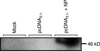
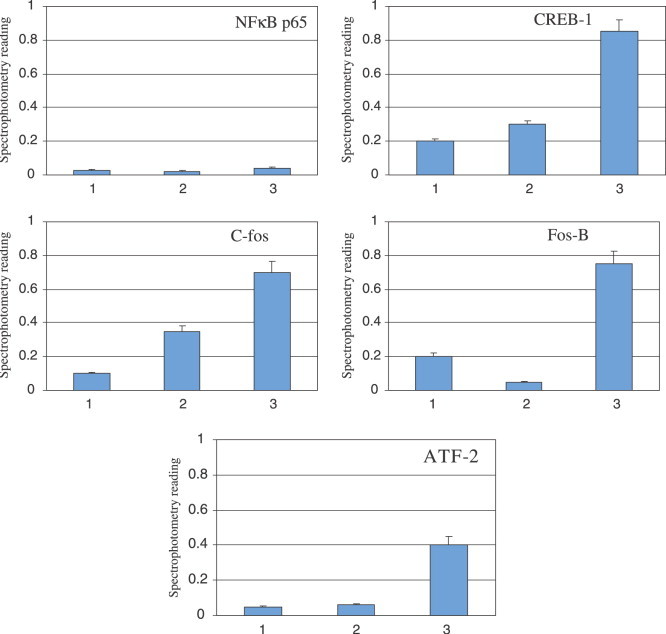
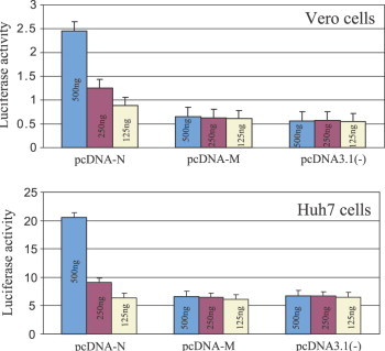
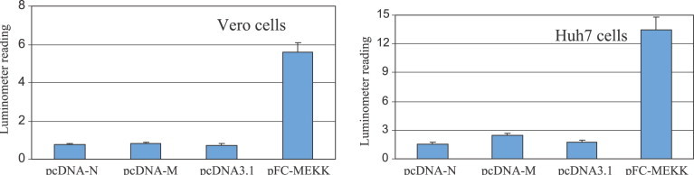
In March 2003, a novel coronavirus was isolated from patients exhibiting atypical pneumonia and subsequently proven to be the causative agent of the disease now referred to as severe acute respiratory syndrome (SARS). The complete genome of the SARS coronavirus (SARS-CoV) has since been sequenced. The SARS-CoV nucleocapsid (SARS-CoV N) shares little homology with other members of the coronavirus family. To determine if the N protein is involved in the regulation of cellular signal transduction, an ELISA-based assay on transcription factors was used. We found that the amount of transcription factors binding to promoter sequences of c-Fos, ATF2, CREB-1, and FosB was increased by the expression of SARS-CoV N. Since these factors are related to AP-1 signal transduction pathway, we investigated whether the AP-1 pathway was activated by SARS-CoV N protein using the PathDetect system. The results demonstrated that the expression of N protein, not the membrane protein (M), activated AP-1 pathway. We also found that SARS-CoV N protein does not activate NF-kappaB pathway, demonstrating that activation of important cellular pathways by SAS-CoV N protein is selective. Thus our data for the first time indicate that SARS-CoV has encoded a strategy to regulate cellular signaling process.
Figures

References
-
- k.V. Holmes, Field’s Virology, vol. I, Lippincott, Williams & Wilkins, Philadelphia, 2001, pp. 1187–1203
-
- Poutanen S.M., Low D.E., Henry B., Finkelstein S., Rose D., Green K., Tellier R., Draker R., Adachi D., Ayers M., Chan A.K., Skowronski D.M., Salit I., Simor A.E., Slutsky A.S., Doyle P.W., Krajden M., Petric M., Brunham R.C., McGeer A.J. Identification of severe acute respiratory syndrome in Canada. N. Engl. J. Med. 2003;348(20):1995–2005. - PubMed
-
- Drosten C., Gunther S., Preiser W., van der Werf S., Brodt H.R., Becker S., Rabenau H., Panning M., Kolesnikova L., Fouchier R.A., Berger A., Burguiere A.M., Cinatl J., Eickmann M., Escriou N., Grywna K., Kramme S., Manuguerra J.C., Muller S., Rickerts V., Sturmer M., Vieth S., Klenk H.D., Osterhaus A.D., Schmitz H., Doerr W. Identification of a novel coronavirus in patients with severe acute respiratory syndrome. N. Engl. J. Med. 2003;348(20):1967–1976. - PubMed
-
- Ksiazek T.G., Erdman D., Goldsmith C.S., Zaki S.R., Peret T., Emery S., Tong S., Urbani C., Comer J.A., Lim W., Rollin P.E., Dowell S.F., Ling A.E., Humphrey C.D., Shieh W.J., Guarner J., Paddock C.D., Rota P., Fields B., DeRisi J., Yang J.Y., Cox N., Hughes J.M., LeDuc J.W., Bellini W.J., Anderson L.J. SARS working group. A novel coronavirus associated with severe acute respiratory syndrome. N. Engl. J. Med. 2003;348(20):1953–1966. - PubMed
-
- Rota P.A., Oberste M.S., Monroe S.S., Nix W.A., Campagnoli R., Icenogle J.P., Penaranda S., Bankamp B., Maher K., Chen M.H., Tong S., Tamin A., Lowe L., Frace M., DeRisi J.L., Chen Q., Wang D., Erdman D.D., Peret T.C., Burns C., Ksiazek T.G., Rollin P.E., Sanchez A., Liffick S., Holloway B., Limor J., McCaustland K., Olsen-Rasmussen M., Fouchier R., Gunther S., Osterhaus A.D., Drosten C., Pallansch M.A., Anderson L.J., Bellini W.J. Characterization of a novel coronavirus associated with severe acute respiratory syndrome. Science. 2003;300(5624):1394–1399. - PubMed
Publication types
MeSH terms
Substances
LinkOut - more resources
Full Text Sources
Miscellaneous
