. 2003 Nov 24;163(4):801-12.
doi: 10.1083/jcb.200304128. Epub 2003 Nov 17.

Smn, the spinal muscular atrophy-determining gene product, modulates axon growth and localization of beta-actin mRNA in growth cones of motoneurons

Affiliations

Smn, the spinal muscular atrophy-determining gene product, modulates axon growth and localization of beta-actin mRNA in growth cones of motoneurons

Wilfried Rossoll et al. J Cell Biol. .

Abstract

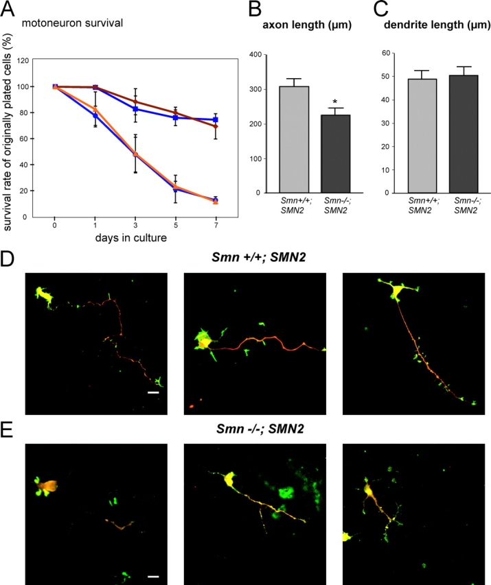
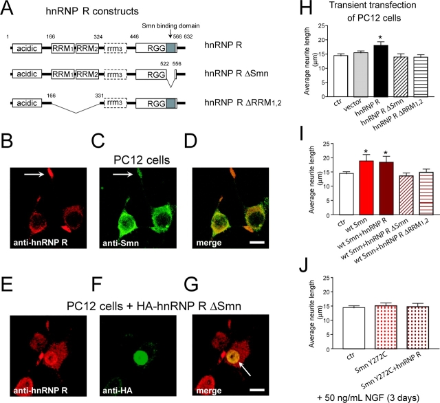
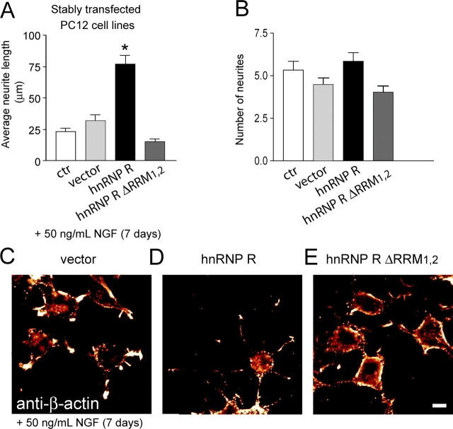
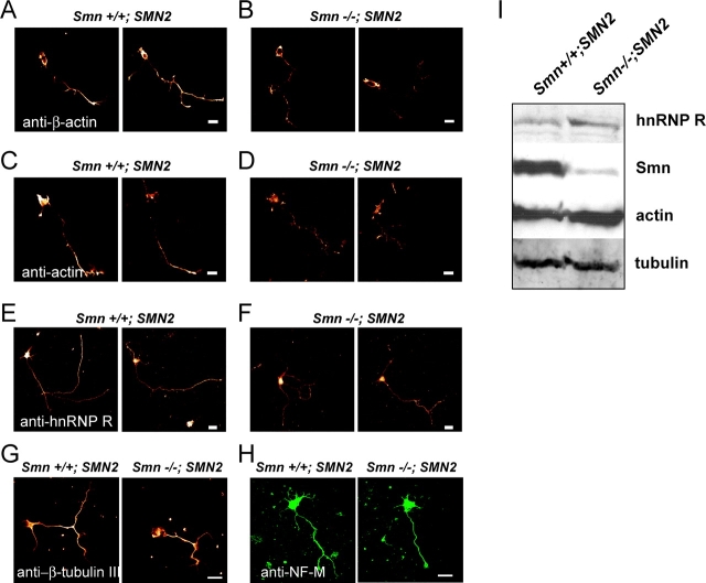
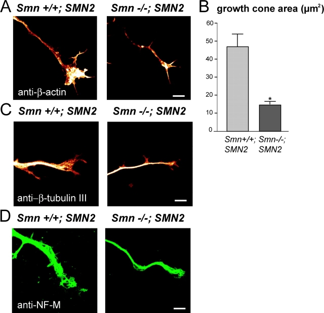
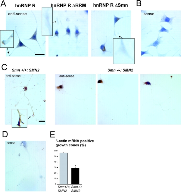
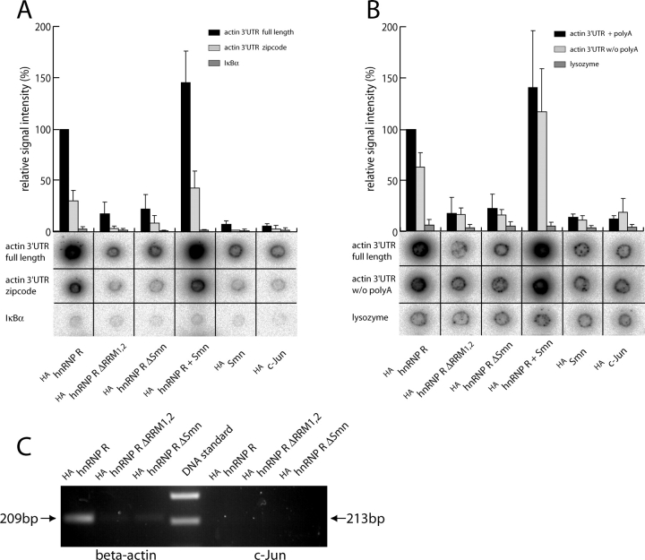
Spinal muscular atrophy (SMA), a common autosomal recessive form of motoneuron disease in infants and young adults, is caused by mutations in the survival motoneuron 1 (SMN1) gene. The corresponding gene product is part of a multiprotein complex involved in the assembly of spliceosomal small nuclear ribonucleoprotein complexes. It is still not understood why reduced levels of the ubiquitously expressed SMN protein specifically cause motoneuron degeneration. Here, we show that motoneurons isolated from an SMA mouse model exhibit normal survival, but reduced axon growth. Overexpression of Smn or its binding partner, heterogeneous nuclear ribonucleoprotein (hnRNP) R, promotes neurite growth in differentiating PC12 cells. Reduced axon growth in Smn-deficient motoneurons correlates with reduced beta-actin protein and mRNA staining in distal axons and growth cones. We also show that hnRNP R associates with the 3' UTR of beta-actin mRNA. Together, these data suggest that a complex of Smn with its binding partner hnRNP R interacts with beta-actin mRNA and translocates to axons and growth cones of motoneurons.


Figures

Figure 1.
Figure 1.
Survival and neurite outgrowth of primary cultured motoneurons from Smn +/+ ; SMN2 and Smn −/− ; SMN2 mice. (A) Survival (percentage of originally plated cells) of Smn +/+ ; SMN2 (blue) and Smn −/− ; SMN2 (orange) motoneurons in the presence (squares) or absence (triangles/circles) of ciliary neurotrophic factor and brain-derived neurotrophic factor. Motoneurons were cultured for 7 d, and survival was scored every 2 d (n = 3). Average length of longest axonal branches (B) and dendritic processes (C) of motoneurons after 5 d in culture is shown. Motoneurons from Smn −/− ; SMN2 mice exhibit a significant reduction in axon length (224.7 ± 20.5 μm vs. 307.6 ± 23.1 μm), whereas dendrite length was not affected. Asterisk denotes significant differences (P < 0.05; n = 4). (D and E) Immunostaining of fixed cells with antibodies against MAP-2 protein (green processes) and axon-specific phospho-tau protein (red processes). Typical examples are shown for Smn +/+ ; SMN2 (D) and Smn −/− ; SMN2 (E) motoneurons. Bar, 20 μm.
Figure 2.
Figure 2.
hnRNP R overexpression increases neurite outgrowth. (A) Schematic representation of the domain structure of wild-type hnRNP R and mutants used to transfect PC12 cells. Either the Smn-binding domain (hnRNP R ΔSmn) or the RNA recognition motifs 1 and 2 (hnRNP R ΔRRM1,2) were deleted. Acidic, protein domain rich in acidic amino acids; RRM, RNA recognition motif; RGG, arginine- glycine-glycine–rich domain. The dashed box indicates a potential additional RRM. Numbers refer to amino acids. (B) PC12 cells were allowed to differentiate for 3 d with NGF, and were then stained with anti-hnRNP R. Intense labeling of growth cones (arrow) and a punctuate nuclear and cytoplasmic staining was observed. (C) anti-Smn staining shows a strong nuclear signal in gem-like structures and a diffuse signal in the cytoplasm and neural processes, including growth cones (arrow). (D) A merge of B and C shows partial colocalization of hnRNP R and Smn in the cell body and colocalization at the growth cones. Bar, 10 μm. In contrast to the endogenous protein (cytoplasmic staining in E and G), most of the HA-tagged protein lacking the Smn-binding domain is localized to the nucleus (nuclear staining in E–G; arrow in G). Bar, 10 μm. After differentiation for 3 d with NGF (H), cells transfected with wild-type hnRNP R show a 25% increase in neurite length (measured as described in the Materials and methods section), whereas overexpression of mutant constructs had no effect on neurite length. (I) Overexpression of wild-type Smn also leads to an increase in neurite extension (∼30%), but cotransfection of wild-type Smn and hnRNP R had no additive effect on neurite extension. Overexpression of mutant hnRNP R with wild-type Smn (I) or mutant SmnY272C with wild-type hnRNP R (J) suppressed the stimulation of neurite outgrowth. Results represent the mean ± SEM of pooled data from four independent transient transfection experiments (H–J). *, P < 0.05 compared with control cells by t test. Only transfected cells as identified by staining with the HA antibody (hnRNP R constructs) or FLAG antibody (Smn constructs) were scored.
Figure 3.
Figure 3.
hnRNP R–overexpressing cell lines exhibit increased neurite outgrowth, whereas hnRNP R ΔRRM1,2 expression leads to reduced β-actin in growth cones. (A) PC12 cell lines overexpressing the indicated constructs were treated for 7 d with NGF. In hnRNP R–overexpressing cell lines, neurite length was increased 2.4-fold (measured as described in the Materials and methods section). (B) The number of neurites was not significantly altered. (C–E) β-Actin immunostaining shows a distinct signal in growth cones of control cell lines (C). hnRNP R–overexpressing cell lines exhibit strong staining in growth cones, which correlates with enhanced neurite growth (D). In hnRNP R ΔRRM1,2–expressing clones (E), β-actin staining was reduced in growth cones and apparently concentrated within cell bodies. In these cells, neurite growth was low. Bar, 10 μm. Results represent the mean ± SEM of pooled data from three different stable cell lines (A and B). *, P < 0.05 between overexpressing cell lines and controls.
Figure 4.
Figure 4.
Actin distribution in axons of primary cultured motoneurons from Sm n −/− ; SMN2 and Smn +/+ ; SMN2 mice. (A and B) Motoneurons from Smn +/+ ; SMN2 and Smn −/− ; SMN2 kept in culture for 5 d were immunostained with β-actin antibody. Motoneurons isolated from Smn +/+ ; SMN2 mice, but not their Smn −/− ; SMN2 littermates, show strong accumulation of β-actin in distal axons and growth cones. (C and D) Immunostaining with pan-actin antibodies. Smn +/+ ; SMN2 motoneurons exhibit a distinct gradient from the shaft to the growth cone and an accumulation of actin in the growth cone, whereas in cells from Smn −/− ; SMN2 littermates, actin staining is predominantly concentrated in cell bodies. (E and F) Immunostaining with anti-hnRNP R antiserum. Control motoneurons show strong staining in the nucleus and a distinct gradient from the shaft to the growth cone, which appears weaker in Smn −/− ; SMN2 cells. (G and H) Immunostaining with anti-tubulin (β-tubulin III) and anti-neurofilament (NF-M) antibodies. There is no difference in the distribution of both cytoskeletal proteins in axons of Smn-deficient and control motoneurons. Bars, 20 μm. (I) Western blot analysis of Smn and hnRNP R in brain extracts from Smn −/− ; SMN2 and control embryonic day 14 embryos. Smn protein is strongly reduced in Smn −/− ; SMN2, whereas hnRNP-R levels are not affected. The blot was subsequently stained for actin, and tubulin was used as a loading control.
Figure 5.
Figure 5.
Reduced growth cone size in primary cultured motoneurons from Smn −/− ; SMN2 mice. (A) High power magnification of β-actin–stained axonal growth cones in cultured Smn +/+ ; SMN2 motoneurons. Bar, 5 μm. (B) Motoneurons from Smn +/+ ; SMN2 (n = 35) and Smn −/− ; SMN2 (n = 31) mice were cultured for 5 d, stained with β-actin, and the area covered by the axonal growth cone was measured. Smn −/− ; SMN2 mice show a significant reduction of the average growth cone area (14.36 ± 2.07 μm2 vs. 46.73 ± 6.9 μm2). (C) β-Tubulin staining and (D) neurofilament (NF-M) staining of axonal growth cones in cultured motoneurons from Smn −/− ; SMN2 and control mice. Bars, 5 μm.
Figure 6.
Figure 6.
Localization of actin mRNA in neurites of differentiated PC12 cells and motoneurons. (A) In situ hybridization with an antisense probe against actin mRNA in differentiated stably transfected PC12 cell lines. Actin mRNA in growth cones is visible as a brown precipitate in cell lines overexpressing wild-type hnRNP R (inset), but not in those overexpressing hnRNP R ΔRRM or hnRNP R ΔSmn. (B) In situ hybridization with an actin sense control in differentiated PC12 cells. (C) In situ hybridization with an antisense probe against actin mRNA in motoneurons derived from Smn +/+ ; SMN2 and Smn −/− ; SMN2 mice shows a reduced accumulation of β-actin mRNA in the distal part of the axons in Smn-deficient cells. (D) An actin sense probe was used as a control and did not reveal a detectable signal in the same experiment. (E) Quantification of the actin mRNA levels in the growth cones by visual scoring. Growth cones from Smn +/+ ; SMN2 (n = 403) and Smn −/− ; SMN2 (n = 368) motoneurons were compared. Smn-deficient motoneurons show a significant reduction in the percentage of actin mRNA–positive growth cones (56.25 ± 1.493 vs. 29.50 ± 1.709). Results represent the mean ± SEM of pooled data from four independent experiments. *, P < 0.05 between control and Smn-deficient motoneurons. Bars, 20 μm.
Figure 7.
Figure 7.
Full-length hnRNP R associates with the 3UTR of β-actin mRNA. HEK 293 cells were transfected with expression vectors as indicated, and HA-tagged proteins were immunoprecipitated 48 h after transfection. (A) Radioactively labeled RNA transcribed from the full-length 3′ UTR of β-actin, the zipcode region of the 3′ UTR, and the IκBα gene was incubated with the immune complexes in solution. After several washes, immunoprecipitates of proteins and bound RNA were spotted on filters for quantification by autoradiography as shown at the bottom. The bar graph indicates the relative amounts of bound radioactive RNA measured by phosphorimaging. Signal intensity for full-length β-actin 3′ UTR bound to full-length hnRNP R was set as 100%. Values are given as relative signal intensities. The full-length 3′ UTR of β-actin and also the zipcode region are bound by full-length hnRNP R, but not by mutant hnRNP R or Smn alone. (B) Radioactively labeled RNA transcribed from the full-length 3′ UTR of β-actin either with or without a poly(A)+ tail and the lysozyme mRNA was incubated with the immune complexes. The graph indicates the amounts of bound radioactively labeled RNA. The 3′ UTR of β-actin both with or without a poly(A)+ tail are bound by full-length hnRNP R. (C) RT-PCR of β-actin coimmunoprecipitated with hnRNP R. HA-tagged wild-type and mutant hnRNP R was immunopurified from differentiated PC12 cell lines with HA-antibody. RNA was isolated from immunoprecipitates, reverse transcribed into cDNA, and RT-PCR was performed with β-actin–specific primers (35 cycles). A clear band of 209 bp can be amplified from RNA bound to wild-type hnRNP R (contamination with genomic DNA would yield a larger fragment including an intron of 123 bp). c-Jun–specific primers were used as a specificity control (expected size of 213 bp).

Similar articles

Cited by

References

    1. Bassell, G.J., H. Zhang, A.L. Byrd, A.M. Femino, R.H. Singer, K.L. Taneja, L.M. Lifshitz, I.M. Herman, and K.S. Kosik. 1998. Sorting of β-actin mRNA and protein to neurites and growth cones in culture. J. Neurosci. 18:251–265. - PMC - PubMed
    1. Bechade, C., P. Rostaing, C. Cisterni, R. Kalisch, V. La Bella, B. Pettmann, and A. Triller. 1999. Subcellular distribution of survival motor neuron (SMN) protein: possible involvement in nucleocytoplasmic and dendritic transport. Eur. J. Neurosci. 11:293–304. - PubMed
    1. Blanc, V., N. Navaratnam, J.O. Henderson, S. Anant, S. Kennedy, A. Jarmuz, J. Scott, and N.O. Davidson. 2001. Identification of GRY-RBP as an apolipoprotein B RNA-binding protein that interacts with both apobec-1 and apobec-1 complementation factor to modulate C to U editing. J. Biol. Chem. 276:10272–10283. - PubMed
    1. Bloom, O., E. Evergren, N. Tomilin, O. Kjaerulff, P. Low, L. Brodin, V.A. Pieribone, P. Greengard, and O. Shupliakov. 2003. Colocalization of synapsin and actin during synaptic vesicle recycling. J. Cell Biol. 161:737–747. - PMC - PubMed
    1. Bommel, H., G. Xie, W. Rossoll, S. Wiese, S. Jablonka, T. Boehm, and M. Sendtner. 2002. Missense mutation in the tubulin-specific chaperone E (Tbce) gene in the mouse mutant progressive motor neuronopathy, a model of human motoneuron disease. J. Cell Biol. 159:563–569. - PMC - PubMed

Publication types

MeSH terms