. 2003 Nov 17;198(10):1609-19.
doi: 10.1084/jem.20030357.

A fail-safe mechanism for negative selection of isotype-switched B cell precursors is regulated by the Fas/FasL pathway

Affiliations

A fail-safe mechanism for negative selection of isotype-switched B cell precursors is regulated by the Fas/FasL pathway

Jane Seagal et al. J Exp Med. .

Abstract

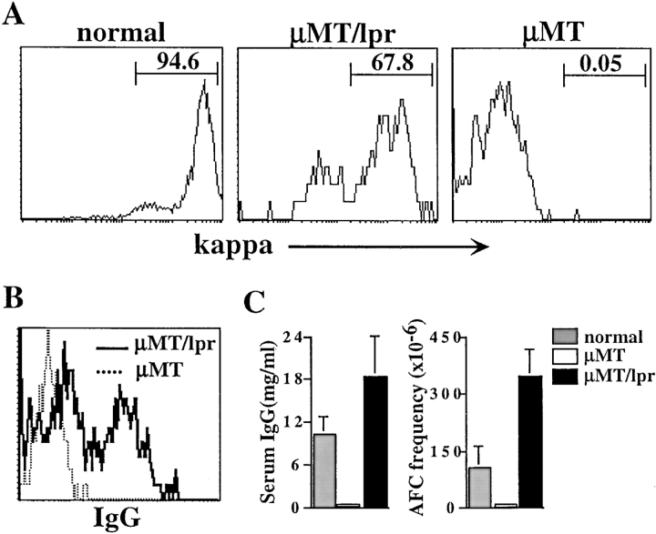
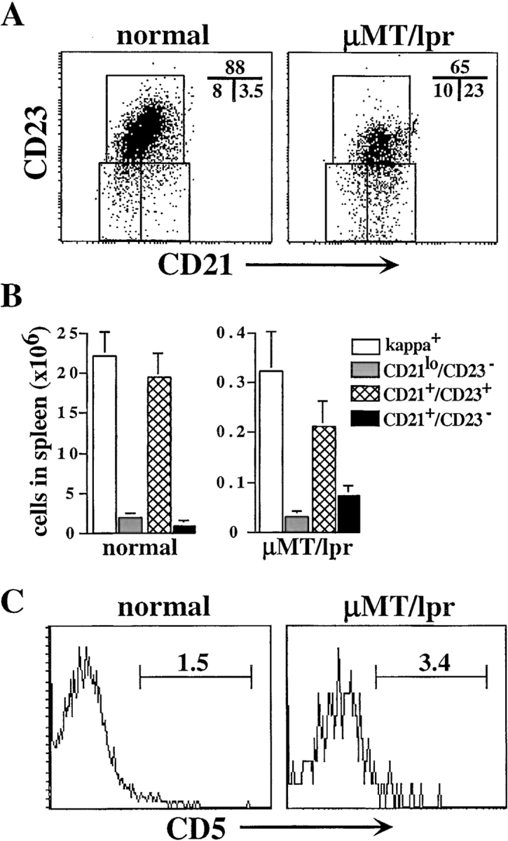
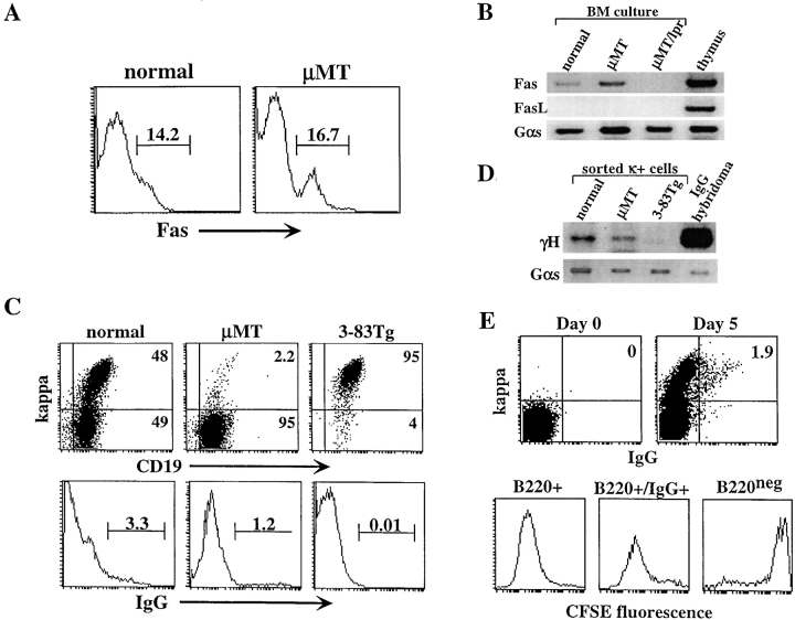
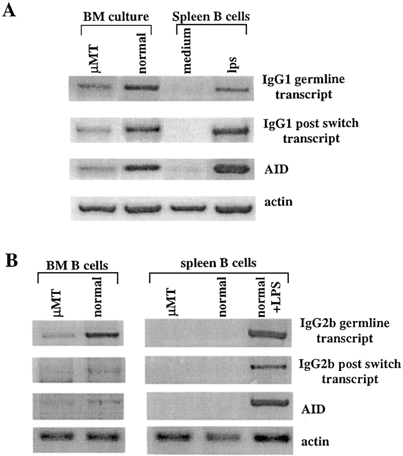
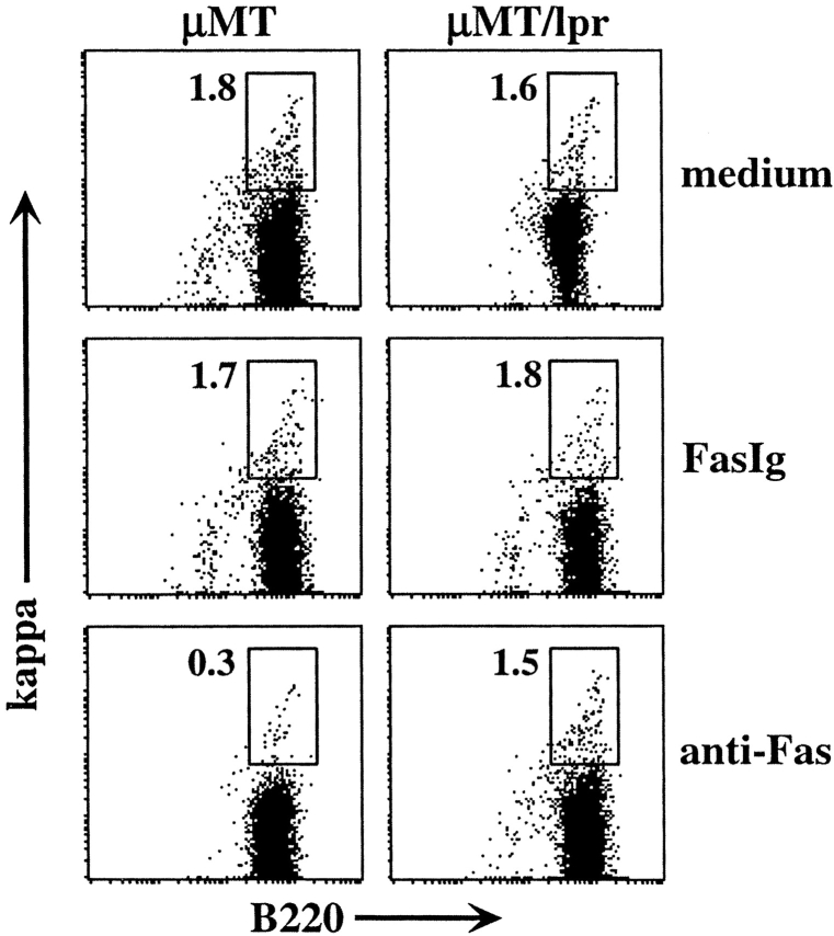
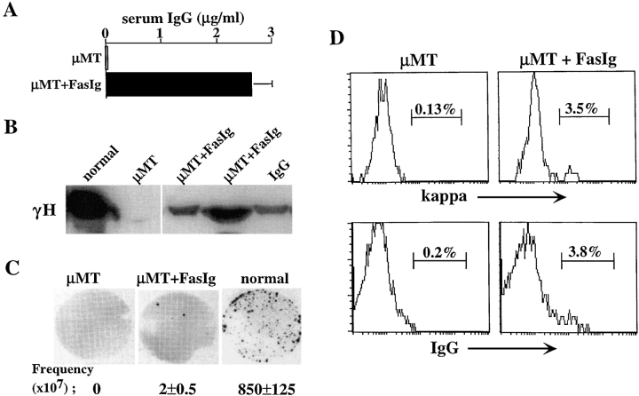
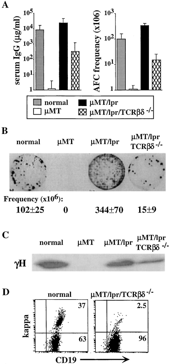
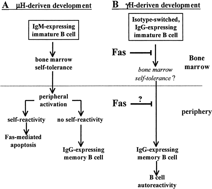
In B lymphocytes, immunoglobulin (Ig)M receptors drive development and construction of naive repertoire, whereas IgG receptors promote formation of the memory B cell compartment. This isotype switching process requires appropriate B cell activation and T cell help. In the absence of T cell help, activated B cells undergo Fas-mediated apoptosis, a peripheral mechanism contributing to the establishment of self-tolerance. Using Igmicro-deficient microMT mouse model, where B cell development is blocked at pro-B stage, here we show an alternative developmental pathway used by isotype-switched B cell precursors. We find that isotype switching occurs normally in B cell precursors and is T independent. Ongoing isotype switching was found in both normal and microMT B cell development as reflected by detection of IgG1 germline and postswitch transcripts as well as activation-induced cytidine deaminase expression, resulting in the generation of IgG-expressing cells. These isotype-switched B cells are negatively selected by Fas pathway, as blocking the Fas/FasL interaction rescues the development of isotype-switched B cells in vivo and in vitro. Similar to memory B cells, isotype-switched B cells have a marginal zone phenotype. We suggest a novel developmental pathway used by isotype-switched B cell precursors that effectively circumvents peripheral tolerance requirements. This developmental pathway, however, is strictly controlled by Fas/FasL interaction to prevent B cell autoimmunity.


Figures

Figure 1.
Figure 1.
Lack of Fas rescues development of isotype-switched B cells in μMT mice. (A) Spleen cells from normal, μMT/lpr, and μMT mice were stained for CD19, B220, and κ. Analysis for κ expression was performed on gated B220+/CD19+ cells. (B) Surface expression of IgG. Spleen cells from the indicated mice were stained for CD19, B220, and IgG. Gated B220+/CD19+ cells were analyzed for IgG expression. (C) Serum IgG and frequency of IgG-producing cells in the indicated mice. Serum samples were collected from mice at 8–12 wk old, and titers of IgG (left) were determined by ELISA. Results shown as mean ± SEM of at least five mice in each group. Numbers of IgG-producing cells (right) were determined by ELISPOT assay and are expressed as frequency of IgG-producing cells per 106 spleen cells. Results are mean ± SEM of at least five mice in each group.
Figure 2.
Figure 2.
Accumulation of isotype-switched B cells in MZ of spleen in μMT/lpr mice. (A) Spleen cells from normal and μMT/lpr mice were stained for CD21, CD23, and κ. FACS® analysis for expression of CD21 and CD23 was performed on gated κ1 cells. (B) Absolute numbers of newly formed (CD21lo/CD23), follicular (CD21+/CD23+), and MZ (CD21+/CD23). Results are mean ± SEM of three mice in each group. (C) FACS® analysis for CD5 expression in gated CD19+1 spleen cells from the indicated mice.
Figure 3.
Figure 3.
Lack of FasL allows development of isotype-switched B cell precursors in vitro. BM cells from normal and μMT mice were cultured for 5 d in the presence of IL-7. (A) Cells grown in culture were stained for B220 and Fas receptor and analyzed by FACS®. Expression of Fas was determined in gated B220+ cells. (B) RNA samples from normal μMT and μMT/lpr BM cultures were analyzed by RT-PCR for Fas and FasL gene expression and for Gαs internal control. Thymus RNA sample was used for positive control. In μMT/lpr sample, no product was observed in the PCR for Fas because oligonucleotides used flank the deleted DNA fragment that is found in lpr mice. (C) Surface expression of κ and IgG in BM cultures. BM cells from normal, μMT, and 3-83μδ Tg mice were cultured in vitro, and cells were stained for B220, CD19, κ, and IgG. Analysis for κ (top) and surface IgG expression was performed on 5,000 gated B220+ cells. (D) RT-PCR amplification for detection of γH expression in BM cultures. κ-expressing cells from the indicated BM cultures were sorted using magnetic beads. mRNA samples from the sorted cells were subjected to RT-PCR amplification using oligonucleotides specific for VHJ558 and γCH1. mRNA from IgG-producing hybridoma was used as control. (E) Normal BM cells were depleted of Ig+ cells using magnetic beads, labeled with CFSE on day 0, washed, and cultured for 5 d with IL-7. Cells were stained and analyzed for IgG and κ coexpression on days 0 and 5 (top). Cells grown in the culture were stained for B220 and IgG and analyzed for CFSE fluorescence (bottom). Analysis was performed on gated B220+ cells (left), B220+/IgG+ cells (middle), or B220 cells (right). Results are representative of three different experiments.
Figure 4.
Figure 4.
Ongoing isotype switching in normal and μMT BM cultures and BM in vivo. (A) BM cells from normal and μMT mouse were cultured in vitro for 5 d in the presence of IL-7. mRNA samples from the cultured cells were subjected to RT-PCR amplification for IgG1 germline and postswitch transcript as well as for AID expression. Normal splenic cells either unstimulated or stimulated with LPS were used as negative and positive controls, respectively. The results shown are representative of three to five different experiments and a total of five to eight mice in each group. (B) B220+ cells were sorted directly from BM or spleen of the indicated mice and mRNA samples were subjected to RT-PCR amplification for AID and IgG2b germline and postswitch transcripts. Representative blots from three mice in each group are shown.
Figure 5.
Figure 5.
Fas receptor ligation eliminates Isotype-switched B cell precursors in vitro. BM cells from μMT and μMT/lpr mice were cultured for a total of 5 d in the presence of IL-7. During the last 48 h, cultured cells were treated with 25 μg/ml FasIg or 2.5 μg/ml anti-Fas antibodies. Cultured cells were stained for CD19, B220, and κ. FACS® analysis was gated to viable CD19+ cells. The plots shown are representative of three different experiments.
Figure 6.
Figure 6.
Blocking the Fas/FasL interaction in vivo rescues isotype-switched B cells in μMT mice. A neutralizing recombinant FasIg protein was purified and engineered into slow releasing PLGA microcapsules. The microcapsules were administered to μMT mice by intramuscular injection of 10 mg/mouse every 10 d for 8 wk. Mice were first injected at 2 wk of age. Control μMT mice were injected with empty microcapsules. 10 d after the last injection, mice were killed. (A) Serum samples from the mice were assayed for the presence of IgG by ELISA. Results are mean ± SEM of three mice in each group. (B) Detection of γH chain in serum samples by Western blotting. Normal serum sample dilution is 1:10. Samples of μMT mice (control and FasIg-treated) were not diluted. Purified IgG was used as control. Line indicates where irrelevant lane was removed digitally. (C) Quantitation of IgG-producing cells in spleens of μMT mice treated with FasIg relative to μMT and normal mice by ELISPOT. Representative ELISPOT membranes and the calculated frequency of IgG-producing cells per 107 spleen cells are shown. Results are mean ± SEM of three mice in each group. (D) FACS® analysis for κ and IgG expression. Spleen cells from the treated mice were stained for CD19, B220, κ, and IgG. Analysis for κ and IgG expression was performed on 10,000 gated CD19+/B220+ cells. The results shown are representative of eight injected mice in three different experiments.
Figure 7.
Figure 7.
Development of isotype-switched B cell precursors is T cell independent. Normal, μMT, μMT/lpr, and μMT/lpr/TCRβδ−/− mice were analyzed for development of isotype-switched B cells and production of serum IgG. (A) Titers of serum IgG were measured by ELISA (left) and are expressed as milligram/milliliter. Frequency of IgG-producing cells in spleens of these mice was quantitated by ELISPOT. The results are mean ± SEM of at least five mice in each group. (B) Representative ELISPOT membrane. Frequencies of plasma cells are mean ± SEM of at least five mice in each group. (C) Detection of γH chain in serum samples by Western blotting. Normal serum sample dilution is 1:10 and μMT/lpr serum dilution is 1:20. Samples of μMT and μMT/lpr/TCRβδ−/− are not diluted. (D) Expression of κ-expressing cells in μMT/lpr/TCRβδ−/− BM cultures. Cells grown in BM cultures prepared from the indicated mice were stained for B220, CD19, and κ. Analysis for κ expression is performed on 5,000 gated B220+ cells.
Figure 8.
Figure 8.
Suggested models for alternative developmental pathways driven by different Ig heavy chain isotypes. The prevailing view of B cell development is shown in A. Most of B cell development is driven by μH chain, and IgM-expressing immature B cells are subjected to self-tolerance in the BM. Mature peripheral B cells express IgM and IgD and become activated upon encountering antigen. Published data suggest that peripheral activation is an important checkpoint to extinguish self-reactivity, mediated by lack of T cell help. In contrast, appropriate T cell help (such as CD40L expression) promotes high affinity, nonself– B cells to develop in the germinal center and undergo isotype switching and differentiate into the memory compartment. The alternative developmental pathway of isotype-switched B cell precursors is shown in B. According to this model, IgG receptor drives IgG-expressing B cells directly into the memory compartment. Because such development circumvents the requirement for peripheral activation, it may confer severe autoimmune risk. In the normal (non-lpr) mouse, such cells are negatively selected by the Fas pathway to prevent their development into the memory compartment and participation in an autoimmune response. Thus, the Fas/FasL pathway is a fail-safe mechanism for elimination of isotype-switched B cell precursors and has a major role in controlling autoimmunity during BM development.

Similar articles

Cited by

References

    1. Rajewsky, K. 1996. Clonal selection and learning in the antibody system. Nature. 381:751–758. - PubMed
    1. Klinman, N.R. 1997. The cellular origins of memory B cells. Semin. Immunol. 9:241–247. - PubMed
    1. Martin, S.W., and C.C. Goodnow. 2002. Burst-enhancing role of the IgG membrane tail as a molecular determinant of memory. Nat. Immunol. 3:182–188. - PubMed
    1. Ahmed, R., and D. Gray. 1996. Immunological memory and protective immunity: understanding their relation. Science. 272:54–60. - PubMed
    1. Han, S., B. Zheng, Y. Takahashi, and G. Kelsoe. 1997. Distinctive characteristics of germinal center B cells. Semin. Immunol. 9:255–260. - PubMed

Publication types