. 2003 Nov 25;100(24):14368-73.
doi: 10.1073/pnas.2336098100. Epub 2003 Nov 17.

Time-restricted role for dendritic activation of the mTOR-p70S6K pathway in the induction of late-phase long-term potentiation in the CA1

Affiliations

Time-restricted role for dendritic activation of the mTOR-p70S6K pathway in the induction of late-phase long-term potentiation in the CA1

Maurizio Cammalleri et al. Proc Natl Acad Sci U S A. .

Abstract

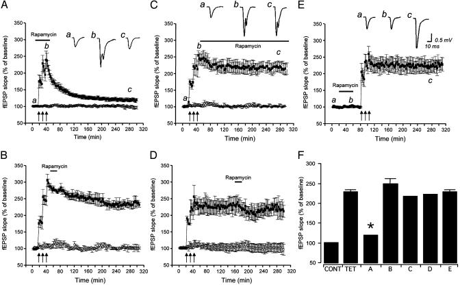
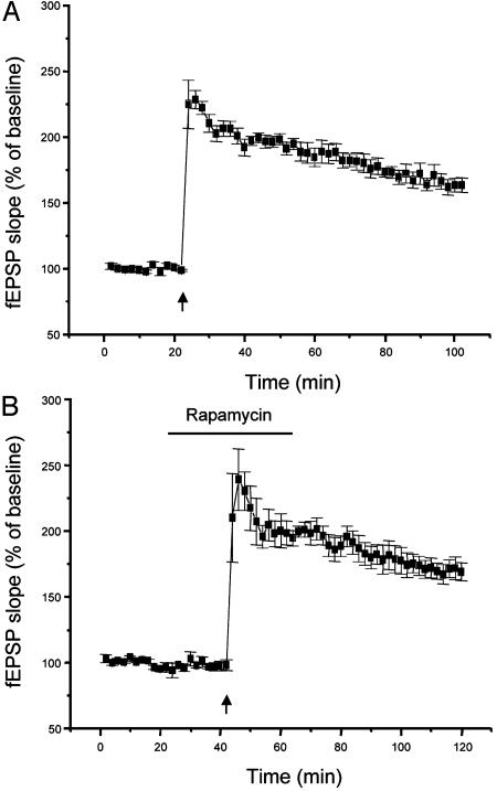
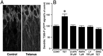
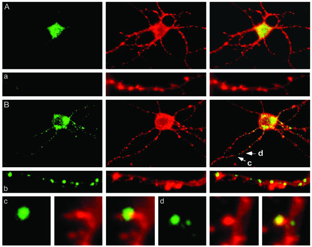
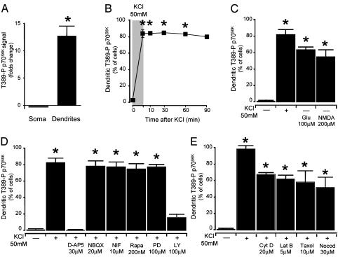
Mammalian target of rapamycin (mTOR) is a key regulator of translational capacity. The mTOR inhibitor rapamycin can prevent forms of protein synthesis-dependent synaptic plasticity such as long-term facilitation in Aplysia and late-phase long-term potentiation (L-LTP) in the hippocampal CA1 region of rodents. In the latter model, two issues remain to be addressed: defining the L-LTP phase sensitive to rapamycin and identifying the site of rapamycin-sensitive protein synthesis. Here, we show that L-LTP is sensitive to application of rapamycin only during the induction paradigm, whereas rapamycin application after the establishment of L-LTP was ineffective. Second, we observed that Thr-389-phosphorylated p70 S6 kinase (p70S6K), the main active phosphoform of the mTOR effector p70S6K, was induced in an N-methyl-D-aspartate and phosphatidylinositol 3-kinase-dependent manner throughout the dendrites but not in the cell bodies of CA1 neurons in hippocampal slices after L-LTP induction. A similar dendrite-wide activation of p70S6K was induced in primary hippocampal neurons by depolarization with KCL or glutamate. In primary hippocampal neurons, the sites of dendritic activation of p70S6K appeared as discrete compartments along dendritic shafts like the hotspots for fast dendritic translation. Conversely, only a subset of dendritic spines also displayed activated p70S6K. Taken together, the present data suggest that the N-methyl-d-aspartate-, phosphatidylinositol 3-kinase-dependent dendritic activation of the mTOR-p70S6K pathway is necessary for the induction phase of protein synthesis-dependent synaptic plasticity. Newly synthesized proteins in dendritic shafts could be targeted selectively to activity-tagged synapses. Thus, coordinated activation of dendrite-wide translation and synaptic-specific activation is likely to be necessary for long-term synaptic plasticity.


Figures

Fig. 1.
Fig. 1.
The mTOR inhibitor rapamycin prevents L-LTP only when applied during induction. fEPSP at Schaffer collateral/commissural fiber-CA1 synapses were simultaneously monitored in two independent pathways (▪, tetanized pathway; ○, untetanized pathway). (A) When rapamycin (1 μM) was transiently (40 min) applied during L-LTP induction, a dramatic decay of fEPSPs was seen (n = 6). A transient rapamycin application 5 min after the delivery of the L-LTP tetanization paradigm (B) or 2 h after completion of the L-LTP induction paradigm (C) did not affect fEPSP slopes (n = 6), nor did a continuous incubation with rapamycin starting 5 min after tetanization (D). (E) L-LTP could be induced after a 40-min rapamycin application and washout, suggesting that the action of rapamycin was reversible. These observations suggest that rapamycin sensitivity of L-LTP is restricted to the induction phase. (F) fEPSP slopes as a percentage of baseline fEPSP in the control untetanized pathway (CONT), untreated tetanized slices (TET), and the conditions displayed in A-E. Measurements of fEPSP slopes taken 240-260 min after completion of the tetanization paradigm. Insets are representative traces of extracellular fEPSPs recorded at the times marked by lowercase letters (*, different from TET, B, C, D, and E; P < 0.01, NS from CONT).
Fig. 2.
Fig. 2.
Rapamycin prevents the conversion of STP into LTP by a group I metabotropic glutamate receptor agonist. (A) Delivery of a tetanic stimulation (50 Hz, for a duration of 0.5 s) subthreshold for the induction of LTP (left arrow) resulted in an STP of fEPSP slopes that rapidly decayed to basal levels. However, an LTP was seen when the same subthreshold tetanic stimulation (right arrow) was delivered to the slices during a brief bath application of the group I metabotropic glutamate receptor agonist trans-(±)-1-amino-1,3-cyclopentanedicarboxylic acid (50 μM for 8 min), as shown by others (18). (B) Such a conversion of STP into LTP could be prevented by a 20-min application of rapamycin (1 μM) during the tetanization paradigm. In this case, fEPSP slopes decayed to baseline within 1 h.
Fig. 3.
Fig. 3.
The induction of a protein synthesis-independent LTP is not sensitive to rapamycin. (A) An LTP of fEPSP was obtained at Schaffer collateral/ commissural fiber-CA1 synapses with a tetanization paradigm consisting of two trains of 0.5-s duration, each at 100 Hz with an interval of 10 s, which only produces a protein synthesis-independent LTP (11, 23). (B) Application of rapamycin (1 μM) during the delivery of such a tetanization paradigm did not modify the level of potentiation of fEPSP slopes.
Fig. 4.
Fig. 4.
NMDA- and PI3K-dependent dendritic accumulation of an active phosphoform of p70S6K (Thr-389-P p70S6K) in tetanized hippocampal slices. (A) Induction of L-LTP at Schaffer collateral/commissural fiber-CA1 synapses in hippocampal slices resulted in the dendritic activation of p70S6K in CA1 neurons (Right). (Left) Control CA1 neurons are shown. (B) The induction of Thr-389-P p70S6K in CA1 dendrites could be prevented by treatment with the NMDA antagonist D-AP5, the mTOR inhibitor rapamycin (RAPA), or the PI3K inhibitor LY294002 during delivery of the tetanization paradigm. Slices that received a single train of tetanization of 100 Hz for 1 s (SING TET) did not show dendritic activation of p70S6K. Thr-389-P p70S6K immunoreactivity in CA1 somata after L-LTP induction was not significantly different (P > 0.05) from untetanized control slices (not shown) (*, P < 0.01, different from control and all other conditions).
Fig. 5.
Fig. 5.
Dendrite-wide induction of Thr-389-P p70S6K in primary hippocampal neurons. (A) Thr-389-P p70S6K was found predominantly in the soma of primary hippocampal neurons (21 days in vitro) (labeling for Thr-389-P p70S6K with a phosphorylation state-specific rabbit polyclonal antibody is shown in green (Right), counterstaining for actin with phycoerythrin-conjugated phalloidin in red in the middle, and the overlay in yellow (Left). Similar results were obtained with a mouse monoclonal antibody specific for Thr-389-P p70S6K. A representative dendrite is shown in a. (B) Thr-389-P p70S6K appeared in distinct punctae along dendrites after depolarization with KCl. A representative dendrite is shown in b. In dendrites (c and d), Thr-389-P p70S6K was also present in a subset of dendritic spines as revealed by double labeling for actin filaments (c and d as shown in B). The morphology of Thr-389-P p70S6K-immunoreactive puncta in dendritic shafts was reminiscent of the morphology of hotspots of dendritic translation recently demonstrated by Aakalu et al. and Job and Eberwine (3, 4) in similarly cultured primary hippocampal neurons.
Fig. 6.
Fig. 6.
Dendritic activation of Thr-389-P p70S6K is NMDA- and PI3K-dependent. (A) Quantification of immunoreactivity for Thr-389-P p70S6K in primary hippocampal neurons after depolarization with KCl (50 mM), as shown in Fig. 5. The intensity of Thr-389-P p70S6K immunoreactivity was greatly increased in dendrites after depolarization with KCl, whereas it was not significantly reduced in the somata. (B) Thr-389-P p70S6K was persistently activated in the dendrites after withdrawal of KCl. (C) Dendritic accumulation of Thr-389-P p70S6K could also be induced by application of glutamate or NMDA. (D) Accumulation of Thr-389-P p70S6K in dendrites was sensitive to the NMDA antagonist D-AP5 and the PI3K inhibitor LY294002 (NIF, nifedipine; Rapa, rapamycin; PD, PD98059; LY, LY294002). (E) Dendritic activation of Thr-389-P p70S6K was not prevented either by the actin polymerization inhibitors cytochalasin D and Latrunculin B or by the inhibitors of microtubule polymerization taxol and nocodazole (*, P < 0.01 from control).

References

    1. Kandel, E. R. (2001) Science 294, 1030-1038. - PubMed
    1. Tang, S. J., Reis, G., Kang, H., Gingras, A. C., Sonenberg, N. & Schuman, E. M. (2002) Proc. Natl. Acad. Sci. USA 99, 467-472. - PMC - PubMed
    1. Aakalu, G., Smith, W. B., Nguyen, N., Jiang, C. & Schuman, E. M. (2001) Neuron 30, 489-502. - PubMed
    1. Job, C. & Eberwine, J. (2001) Proc. Natl. Acad. Sci. USA 98, 13037-13042. - PMC - PubMed
    1. Pullen, N. & Thomas, G. (1997) FEBS Lett. 410, 78-82. - PubMed

Publication types

MeSH terms

LinkOut - more resources