. 2003 Nov 25;100(24):14439-44.
doi: 10.1073/pnas.2435457100. Epub 2003 Nov 17.

Neuroactive steroids reduce neuronal excitability by selectively enhancing tonic inhibition mediated by delta subunit-containing GABAA receptors

Affiliations

Neuroactive steroids reduce neuronal excitability by selectively enhancing tonic inhibition mediated by delta subunit-containing GABAA receptors

Brandon M Stell et al. Proc Natl Acad Sci U S A. .

Abstract

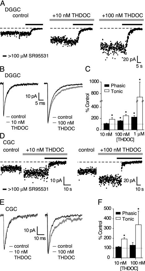
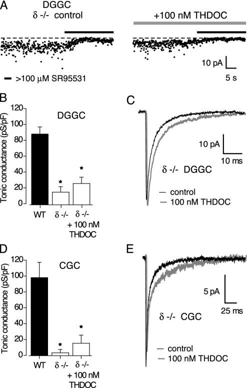
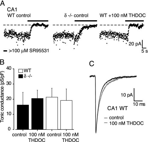
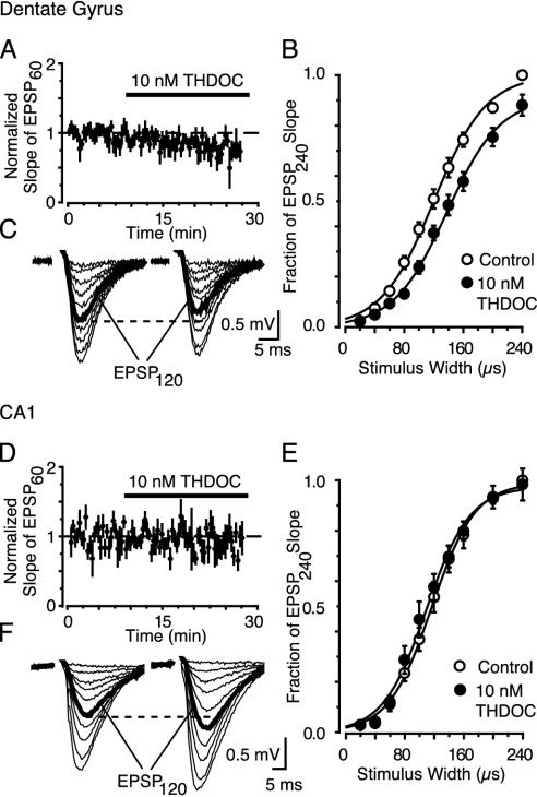
Neuroactive steroids are potent modulators of gamma-aminobutyric acid type A receptors (GABAARs), and their behavioral effects are generally viewed in terms of altered inhibitory synaptic transmission. Here we report that, at concentrations known to occur in vivo, neuroactive steroids specifically enhance a tonic inhibitory conductance in central neurons that is mediated by extrasynaptic delta subunit-containing GABAARs. The neurosteroid-induced augmentation of this tonic conductance decreases neuronal excitability. Fluctuations in the circulating concentrations of endogenous neuroactive steroids have been implicated in the genesis of premenstrual syndrome, postpartum depression, and other anxiety disorders. Recognition that delta subunit-containing GABAARs responsible for a tonic conductance are a preferential target for neuroactive steroids may lead to novel pharmacological approaches for the treatment of these common conditions.


Figures

Fig. 1.
Fig. 1.
Selective modulation by THDOC of a tonic GABAAR-mediated conductance. (A) Current values were averaged over 10-ms epochs at 100-ms intervals, under control conditions and in the presence of 10 and 100 nM THDOC. Horizontal bars indicate the application of the GABAAR antagonist SR95531 (final concentration ≥100 μM). The dotted line is the mean current after complete block of GABAARs used to calculate the magnitude of the tonic GABAAR-mediated conductance. This conductance is increased in the presence of both 10 and 100 nM THDOC, and GABAAR blockade rapidly reduces both its magnitude and variance. (B) Effects of 10 and 100 nM THDOC on averaged sIPSCs recorded in two DGGCs. (C) Concentration-dependent effects of THDOC on the tonic conductance (open bars) and average charge transfer through phasic sIPSCs (filled bars) expressed as a percentage of control values in the absence of THDOC (dashed line). Error bars denote SEM, and asterisks denote significance (P < 0.05). (D) Increase in baseline current in two CGCs by 5-min applications of 10 or 100 nM THDOC and the effect of GABAAR blockade (filled bars). (E and F) Same as B and C for CGCs.
Fig. 2.
Fig. 2.
Reduced tonic conductance in δ-/- mice. (A) Effects of GABAAR blockade on the holding current in two representative DGGCs from δ-/- mice (details as in Fig. 1 A). The tonic current is smaller than that seen in wild-type mice, and is not increased by 100 nM THDOC (see Fig. 1 A). (B) Tonic conductance in DGGCs from wild-type (filled bar) and δ-/- mice (open bars). Note the significant reduction in tonic conductance (asterisk), and the lack of effect of 100 nM THDOC, in δ-/- mice. (C) Averaged sIPSCs recorded in a DGGC from a δ-/- mouse, illustrating the effect of 100 nM THDOC. (D and E) Data from CGCs.
Fig. 3.
Fig. 3.
The tonic conductance in CA1 pyramidal cells is not mediated by δ subunit-containing GABAARs and is not sensitive to 100 nM THDOC. (A) The effects of GABAAR blockade on the holding current in three representative CA1 pyramidal cells from wild-type or δ-/- mice (details as in Fig. 1 A). (B) The tonic conductances in wild-type (filled bars) and δ-/- (open bars) neurons were similar in the absence or presence of 100 nM THDOC. (C) Averaged sIPSCs recorded from a wild-type CA1 pyramidal cell in the absence and presence of 100 nM THDOC. The small prolongation of the sIPSC decay (≈25% increase in τ2) did not affect the average charge transfer (305 ± 38 fC in control; 319 ± 90 fC in THDOC; n = 3) or τw (see Materials and Methods).
Fig. 4.
Fig. 4.
A physiological concentration (10 nM) of THDOC reduces the fEPSP slope when δ-subunits are present. (A and D) Time vs. average fEPSP slope evoked by W = 60 μs (EPSP60). Data were pooled and normalized to the average fEPSP slope evoked during first 10 min (n = 4 slices). (B and E) fEPSPs evoked during stimulus response curves before (Left) and after (Right)10nM THDOC (20 min). The dashed line compares the two EPSP120 responses (bold, evoked by a W closest to the fitted W50). (C and F) Stimulus–response curves after 20 min of 10 nM THDOC (•) in the dentate gyrus (C) or the striatum radiatum of CA1 (F). Data (±SEM) are normalized to the slope of the EPSP240 (evoked by W = 240 μs) under control conditions (○). Lines represent the Boltzman equation fitted to the means.

References

    1. McKernan, R. M. & Whiting, P. J. (1996) Trends Neurosci. 19, 139-143. - PubMed
    1. Majewska, M. D. (1992) Prog. Neurobiol. 38, 379-395. - PubMed
    1. Olsen, R. W. & Sapp, D. W. (1995) Adv. Biochem. Psychopharmacol. 48, 57-74. - PubMed
    1. Paul, S. M. & Purdy, R. H. (1992) FASEB J. 6, 2311-2322. - PubMed
    1. Smith, S. S. (2001) Cell. Mol. Life Sci. 58, 1263-1275. - PMC - PubMed

Publication types

MeSH terms