. 2003 Nov 25;100(24):13755-60.
doi: 10.1073/pnas.2331316100. Epub 2003 Nov 17.

Microfabricated needles for transdermal delivery of macromolecules and nanoparticles: fabrication methods and transport studies

Affiliations

Microfabricated needles for transdermal delivery of macromolecules and nanoparticles: fabrication methods and transport studies

Devin V McAllister et al. Proc Natl Acad Sci U S A. .

Abstract

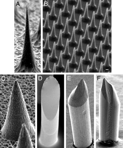
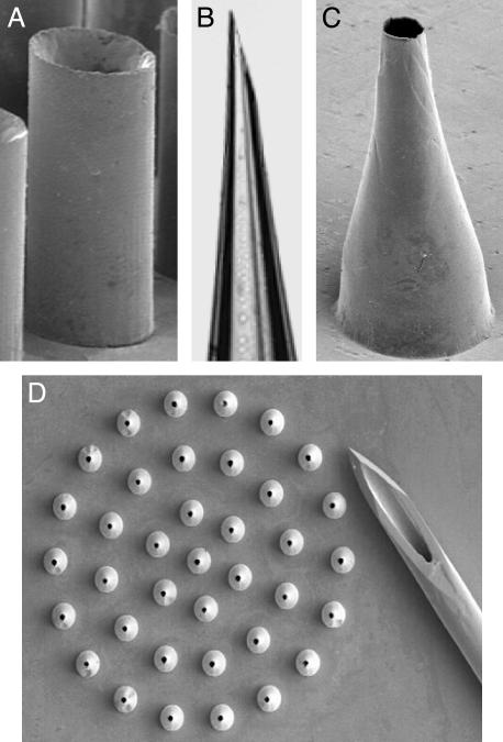
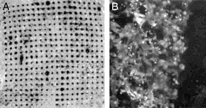
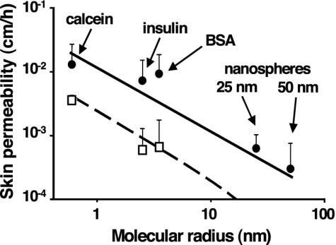
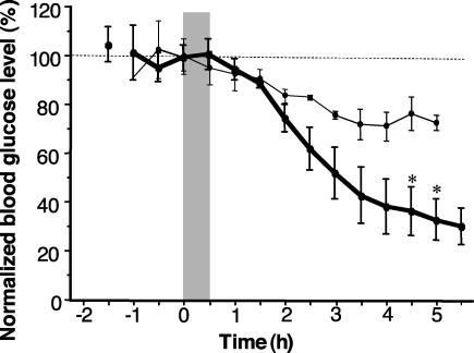
Arrays of micrometer-scale needles could be used to deliver drugs, proteins, and particles across skin in a minimally invasive manner. We therefore developed microfabrication techniques for silicon, metal, and biodegradable polymer microneedle arrays having solid and hollow bores with tapered and beveled tips and feature sizes from 1 to 1,000 microm. When solid microneedles were used, skin permeability was increased in vitro by orders of magnitude for macromolecules and particles up to 50 nm in radius. Intracellular delivery of molecules into viable cells was also achieved with high efficiency. Hollow microneedles permitted flow of microliter quantities into skin in vivo, including microinjection of insulin to reduce blood glucose levels in diabetic rats.


Figures

Fig. 1.
Fig. 1.
Solid microneedles fabricated out of silicon, polymer, and metal, imaged by scanning electron microscopy. (A) Silicon microneedle (150 μm tall) from a 400-needle array etched out of a silicon substrate. (B) Section of an array containing 160,000 silicon microneedles (25 μm tall). (C) Metal microneedle (120 μm tall) from a 400-needle array made by electrodepositing onto a polymeric mold. (D–F) Biodegradable polymer microneedles with beveled tips from 100-needle arrays made by filling polymeric molds. (D) Flat-bevel tip made of polylactic acid (400 μm tall). (E) Curved-bevel tip made of polyglycolic acid (600 μm tall). (F) Curved-bevel tip with a groove etched along the full length of the needle made of polyglycolic acid (400 μm tall).
Fig. 2.
Fig. 2.
Hollow microneedles fabricated out of silicon, metal, and glass imaged by optical and scanning electron microscopy. (A) Straight-walled metal microneedle from a 100-needle array fabricated by electrodeposition onto a polymer mold (200 μm tall). (B) Tip of a tapered, beveled, glass microneedle made by conventional micropipette puller (900-μm length shown). (C) Tapered, metal microneedle (500 μm tall) from a 37-needle array made by electrodeposition onto a polymeric mold. (D) Array of tapered metal microneedles (500 μm height) shown next to the tip of a 26-gauge hypodermic needle.
Fig. 3.
Fig. 3.
Optical micrographs showing molecular transport induced by microneedles. (A) An array of microneedles (Fig. 1 A) was inserted and removed from human cadaver skin, which was subsequently exposed to trypan blue on the exterior stratum corneum side. The array of dark dots shown on the interior viable epidermis side indicates that microneedles created transport pathways across the tissue. (B) An array of 25-μm-tall microneedles (Fig. 1B) was briefly inserted into a monolayer of prostate cancer cells bathed in a solution of calcein, a cell-impermeant fluorescent tracer. Cells on the left were treated with microneedles and show bright fluorescence, indicating uptake of calcein. Cells on the right (dark area) were not treated with microneedles and show little fluorescence, indicating little or no uptake. Most cells remained viable after treatment (data not shown).
Fig. 4.
Fig. 4.
Skin permeability to molecules and particles of different sizes after treatment with microneedles. The permeability of human cadaver epidermis was increased by orders of magnitude with a 400-needle array (Fig. 1 A) inserted (□) and after the array was removed (•) for calcein, insulin, BSA, and latex nanospheres of 25 nm and 50 nm radius. Permeability to nanospheres with needles inserted was below the detection limit, on the order of 10–4 cm/h. In the absence of microneedles, permeability to all compounds was below their detection limits, on the order of 10–6 to 10–4 cm/h (data not shown). Mean values ± SEM are shown for at least six replicates. Predictions are shown for needles inserted (dashed line) and needles removed (solid line) by using a model requiring no adjustable parameters (Eq. 1 coupled with the Stokes–Einstein equation to interrelate molecular radius and diffusivity).
Fig. 5.
Fig. 5.
Blood glucose levels in diabetic hairless rats shown as a function of time after injection of insulin solution at 10 psi (•) (n = 3, 4.3 ± 0.7 μl delivered) or 14 psi (○) (n = 4, 28 ± 11 μl delivered). Blood glucose level was measured before and after insulin was microinjected through a hollow, glass microneedle inserted into rat skin for 30 min (shaded region). Each data point represents the mean (±SEM) of three or four measurements on different rats.

Similar articles

Cited by

References

    1. Madou, M. (1997) Fundamentals of Microfabrication (CRC, Boca Raton, FL).
    1. Chen, J. & Wise, K. D. (1997) IEEE Trans. Biomed. Eng. 44, 760–769. - PubMed
    1. Brazzle, J., Papautsky, I. & Frazier, A. B. (1999) IEEE Eng. Med. Biol. Mag. 18, 53–58. - PubMed
    1. Lin, L. & Pisano, A. P. (1999) IEEE J. Micromech. Syst. 8, 78–84.
    1. Stoeber, B. & Liepmann, D. (2000) Proc. ASME MEMS Dev. 2000 IMECE 2, 355–359.

Publication types

MeSH terms

LinkOut - more resources