. 2003 Nov 25;100(24):14327-32.
doi: 10.1073/pnas.2332326100. Epub 2003 Nov 17.

Apoptosis induced by environmental stresses and amphotericin B in Candida albicans

Affiliations

Apoptosis induced by environmental stresses and amphotericin B in Candida albicans

Andrew J Phillips et al. Proc Natl Acad Sci U S A. .

Abstract

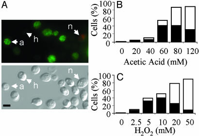
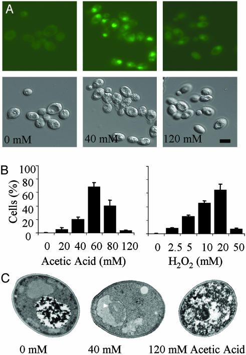
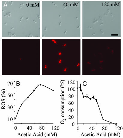
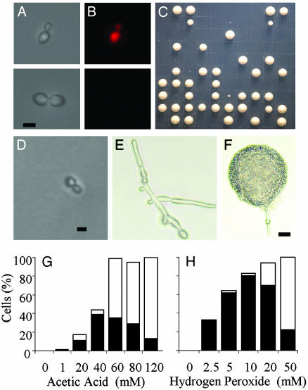
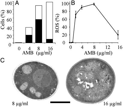
New antifungal agents are urgently required to combat life-threatening infections caused by opportunistic fungal pathogens like Candida albicans. The manipulation of endogenous fungal programmed cell death responses could provide a basis for future therapies. Here we assess the physiology of death in C. albicans in response to environmental stresses (acetic acid and hydrogen peroxide) and an antifungal agent (amphotericin B). Exposure of C. albicans to 40-60 mM acetic acid, 5-10 mM hydrogen peroxide, or 4-8 microg.ml-1 amphotericin B produced cellular changes reminiscent of mammalian apoptosis. Nonviable cells that excluded propidium iodide displayed the apoptotic marker phosphatidylserine (as shown by annexin-V-FITC labeling), were terminal deoxynucleotidyltransferase-mediated dUTP nick end labeling (TUNEL)-positive (indicating nuclease-mediated double-strand DNA breakage), and produced reactive oxygen species. Ultrastructural changes in apoptotic cells included chromatin condensation and margination, separation of the nuclear envelope, and nuclear fragmentation. C. albicans cells treated at higher doses of these compounds showed cellular changes characteristic of necrosis. Necrotic cells displayed reduced TUNEL staining, a lack of surface phosphatidylserine, limited reactive oxygen species production, and an inability to exclude propidium iodide. Necrotic cells lacked defined nuclei and showed extensive intracellular vacuolization. Apoptosis in C. albicans was associated with an accumulation of cells in the G2/M phase of the cell cycle, and under some apoptosis-inducing conditions, significant proportions of yeast cells switched to hyphal growth before dying. This is a demonstration of apoptosis in a medically important fungal pathogen.


Figures

Fig. 1.
Fig. 1.
Exposure of PS in C. albicans.(A) Fluorescence and DIC micrographs showing healthy (h) annexin(-) PI(-) protoplasts, apoptotic (a) annexin(+) PI(-) protoplasts, and necrotic (n) annexin(+) PI(+) protoplasts after treatment with acetic acid. (Scale bar: 5 μm.) (B and C) Percentage of cells that are classified as apoptotic [annexin(+) PI(-); black bars] and necrotic [annexin(+/-) PI(+); white bars] after treatment with acetic acid (B) or hydrogen peroxide (C).
Fig. 2.
Fig. 2.
DNA damage revealed by the TUNEL assay in C. albicans. (A) Fluorescence and DIC micrographs showing TUNEL(+) protoplasts and TUNEL(-) protoplasts after treatment with acetic acid. (Scale bar: 5 μm.) (B) Percentage of cells that contain damaged DNA as revealed by the TUNEL assay after treatment with acetic acid and hydrogen peroxide. (C) Transmission electron micrographs show morphological changes characteristic of apoptosis (chromatin condensation and margination, white arrows) and necrosis (organellar swelling and membrane disintegration, black arrows) in cells treated with different doses of acetic acid. (Scale bar: 5 μm.)
Fig. 3.
Fig. 3.
Apoptotic C. albicans cells produce ROS. (A) Fluorescence and DIC micrographs showing DHR123 staining in cells treated with acetic acid. (Scale bar: 5 μm.) (B) Percentage of cells that contain ROS revealed by DHR staining after treatment with acetic acid for 200 min at 30°C. (C) Oxygen consumption rates of cells treated with acetic acid.
Fig. 4.
Fig. 4.
Single-cell manipulations reveal treatments (40 mM acetic acid shown) that maximize the production of PI(-), nonviable cells (nominally apoptotic). (A and B) Representative micrographs showing individual PI(+) and PI(-) cells after micromanipulation onto a grid. (C) Plate showing colonies after single-cell manipulation. (D-F) Cells that failed to form a colony (D), switched to the production of hyphae and then arrested (E), or switched and then formed a colony (F). (Scale bars: D and E, 5 μm; F, 20 μm.) (G and H) Percentage of apoptotic PI(-), nongrowing cells (black bars) and the percentage of necrotic PI(+), nongrowing cells (white bars).
Fig. 5.
Fig. 5.
C. albicans cells treated with AMB display apoptotic phenotypes. (A) Single-cell manipulation data showing the percentage of apoptotic PI(-), nongrowing cells (black bars) and the percentage of necrotic PI(+), nongrowing cells (white bars) for different doses of AMB. (B) Percentage of cells that contain ROS (revealed by DHR staining) after treatment with AMB. (C) Transmission electron micrographs reveal morphological changes characteristic of apoptosis and necrosis in C. albicans cells treated with AMB. (Scale bar: 5 μm.)

Similar articles

Cited by

References

    1. Odds, F. C. (1988) Candida and Candidosis (Baillière Tindall, London), 2nd Ed.
    1. Banerjee, S. N., Emori, T. G., Culver, D. H., Gaynes, R. P., Jarvis, W. R., Horan, T., Edwards, J. R., Tolson, J., Henderson, T. & Martone, W. J. (1991) Am. J. Med. 91, 86S-89S. - PubMed
    1. Lamagni, T. L., Evans, B. G., Shigematsu, M. & Johnson, E. M. (2001) Epidemiol. Infect. 126, 397-414. - PMC - PubMed
    1. Pfaller, M. A. (1996) Clin. Infect. Dis. 22, S89-S94. - PubMed
    1. Mah, T. F. & O'Toole, G. A. (2001) Trends Microbiol. 9, 34-39. - PubMed

Publication types

MeSH terms