. 2003 Dec;9(12):1464-75.
doi: 10.1261/rna.5290903.

The ability to form full-length intron RNA circles is a general property of nuclear group I introns

Affiliations

The ability to form full-length intron RNA circles is a general property of nuclear group I introns

Henrik Nielsen et al. RNA. 2003 Dec.

Abstract

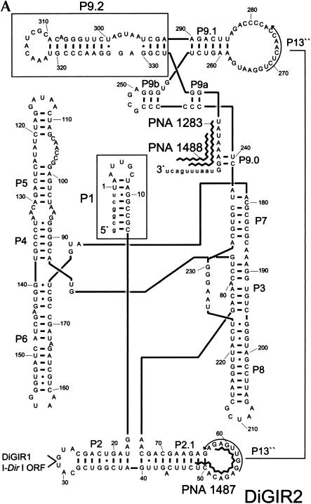
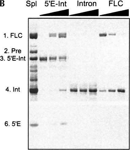
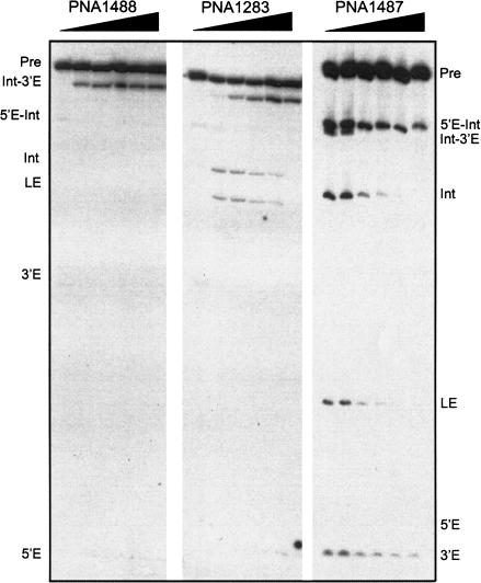
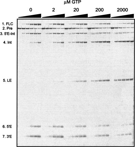
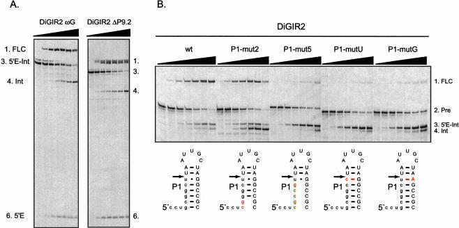
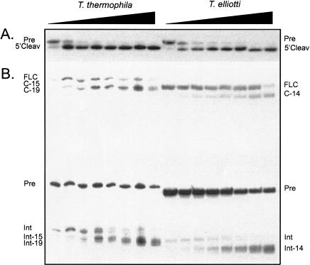
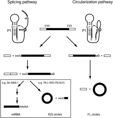
In addition to splicing, group I intron RNA is capable of an alternative two-step processing pathway that results in the formation of full-length intron circular RNA. The circularization pathway is initiated by hydrolytic cleavage at the 3' splice site and followed by a transesterification reaction in which the intron terminal guanosine attacks the 5' splice site presented in a structure analogous to that of the first step of splicing. The products of the reactions are full-length circular intron and unligated exons. For this reason, the circularization reaction is to the benefit of the intron at the expense of the host. The circularization pathway has distinct structural requirements that differ from those of splicing and appears to be specifically suppressed in vivo. The ability to form full-length circles is found in all types of nuclear group I introns, including those from the Tetrahymena ribosomal DNA. The biological function of the full-length circles is not known, but the fact that the circles contain the entire genetic information of the intron suggests a role in intron mobility.


Figures

FIGURE 1.
FIGURE 1.
Structural and hydrolytic features of DiGIR2. (A) Secondary structure model of DiGIR2. Structural segments important in this study (P1 and P9.2) are boxed, and specificities of the peptide nucleic acids PNA1283, PNA1487, and PNA1488 are indicated. (B) Incubation of 3′ SS hydrolyzed precursor RNA generates the full-length circular intron RNA. Individual RNA species were purified from gels and incubated under splicing conditions without GTP for various times (0, 30, and 60 min, indicated by triangles above lanes). RNAs were subsequently separated on an 8 M urea/4% polyacrylamide gel in 0.4× TBE buffer. The RNAs are the 5′-exon-intron RNA (5′E-Int), the intron RNA (Int), and the full-length circular intron RNA (FLC). The circularization junction in FLC was amplified by RT-PCR, sequenced, and verified to correspond to full-length intron circularization (data not shown).
FIGURE 1.
FIGURE 1.
Structural and hydrolytic features of DiGIR2. (A) Secondary structure model of DiGIR2. Structural segments important in this study (P1 and P9.2) are boxed, and specificities of the peptide nucleic acids PNA1283, PNA1487, and PNA1488 are indicated. (B) Incubation of 3′ SS hydrolyzed precursor RNA generates the full-length circular intron RNA. Individual RNA species were purified from gels and incubated under splicing conditions without GTP for various times (0, 30, and 60 min, indicated by triangles above lanes). RNAs were subsequently separated on an 8 M urea/4% polyacrylamide gel in 0.4× TBE buffer. The RNAs are the 5′-exon-intron RNA (5′E-Int), the intron RNA (Int), and the full-length circular intron RNA (FLC). The circularization junction in FLC was amplified by RT-PCR, sequenced, and verified to correspond to full-length intron circularization (data not shown).
FIGURE 2.
FIGURE 2.
PNA inhibition experiments. DiGIR2 templates were transcribed in the presence of PNA1488, PNA1283, or PNA1487 complementary to the 3′ SS or to the L2.1 (see Fig. 1A ▶). Different amounts of PNA were included with the transcription reaction (0, 1, 2, 5, 10, 25 pmole/10 μL) and the transcripts analyzed on 8 M urea/5% polyacrylamide gels. The positions in the gels of the relevant RNA species are indicated. Note that the DiGIR2 used in these experiments has a longer 3′ exon compared to that used in other experiments.
FIGURE 3.
FIGURE 3.
Competition between added exogenous guanosine (GTP) and terminal guanosine (ωG) in DiGIR2 intron splicing and 3′ hydrolysis. The primary transcripts (Pre) were incubated under splicing conditions at varying GTP concentrations (0, 2, 20, 200, and 2000 μM) in time course experiments (0, 2, 5, 15, 30, and 60 min). RNAs were subsequently separated on an 8 M urea/5% polyacrylamide gel. The positions in the gels of the relevant RNA species are indicated. Note that the RNA species slightly smaller than RNA 3 present at early time points and high GTP concentrations corresponds to an Intron-3′-exon (Int-3′E) RNA. The circular species (RNA1) was eluted from the gels and purified. The circularization junction was amplified by RT-PCR and sequenced and found to correspond to a full-length intron circle (data not shown).
FIGURE 4.
FIGURE 4.
Structural requirements in full-length intron circularization. (A) Analysis of circularization of intron transcripts ending exactly in ωG. RNA transcripts corresponding to 5′-exon-intron (5′E-Int) of DiGIR2 and DiGIR2-ΔP9.2 were incubated under splicing conditions in time course experiments (0, 5, 15, 30, 60, 90, and 120 min), and subsequently separated on an 8 M urea/5% polyacrylamide gel. (B) The importance of the P1 segment in full-length circularization of DiGIR2. RNA transcripts corresponding to different P1 mutants were incubated under splicing conditions in time course experiments (0, 2, 5, 10, 20, 30, and 60 min), separated on an 8 M urea/5% polyacrylamide gel, and analyzed for full-length circle formation.
FIGURE 5.
FIGURE 5.
Hydrolysis at the 3′ SS and circularization of Tetrahymena introns. The two steps of the circularization pathway were analyzed independently in homologous introns from two different Tetrahymena species (T. thermophila and T. elliotti). (A) Intron transcripts lacking P1 (and consequently the 5′ SS) were analyzed for the 3′-SS hydrolysis step of the reaction. (B) Intron transcripts that include the 5′ exon, but terminate exactly at the ωG, were analyzed for the circularization step of the reaction. All the experiments were performed as a time-course experiment (0, 2, 5, 10, 15, 30, 60, 120 min) using standard splicing conditions and the RNAs analyzed on either 8 M urea/6% polyacrylamide gels (A) or 8 M urea/4% polyacrylamide gels in 0.4× TBE buffer (B). The positions in the gel of the various RNAs from T. thermophila and T. elliotti are indicated to the left and right, respectively. For example, C-15 and Int-15 refer to circular and linear intron species lacking 15 nt at the 5′ terminal. The identity of the circular species were verified by gel purification, RT-PCR, and sequencing (data not shown).
FIGURE 6.
FIGURE 6.
Schematic representation of the two processing pathways in nuclear group I introns. The splicing pathway (left) is initiated by binding of exogenous guanosine (exoG) to the intron internal G binding site followed by nucleophilic attack at the 5′ SS presented in the P1 context. Two transesterification reactions referred to as the first and second steps of splicing result in a spliced out intron with the exoG attached at the 5′ end and spliced exons. The spliced out intron can undergo one of two different sets of reactions. In twin-ribozyme introns (e.g., Dir.S956-1), a set of reactions leads to the formation of an mRNA encoding a homing endonuclease. In most nuclear group I introns (e.g., Tth.L1925), the ωG attacks an internal phosphodiester bond close to the 5′ end presented in a way analogous to the P1 presentation of the 5′ SS. As a result, shortened circles are formed (IGS circles). The circularization pathway (right) is initiated by hydrolysis at the 3′ SS followed by nucleophilic attack of ωG at the 5′ SS presented in the P1 context. This pathway involves one site-specific hydrolysis reaction and one transesterification and results in the formation of full-length intron circles, leaving the exons unligated.

References

    1. Been, M.D. and Cech, T.R. 1986. One binding site determines sequence specificity of Tetrahymena pre-rRNA self-splicing, trans-splicing, and RNA enzyme activity. Cell 47: 207–216. - PubMed
    1. ———. 1987. Selection of circularization sites in a group I IVS RNA requires multiple alignments of an internal template-like sequence. Cell 50: 951–961. - PubMed
    1. Been, M.D. and Perrotta, A.T. 1991. Group I intron self-splicing with adenosine: Evidence for a single nucleoside-binding site. Science 252: 434–437. - PubMed
    1. Bhattacharya, D. 1998. The origin and evolution of protist group I introns. Protist 149: 113–122. - PubMed
    1. Brehm, S.L. and Cech, T.R. 1983. Fate of an intervening sequence ribonucleic acid: Excision and cyclization of the Tetrahymena ribosomal ribonucleic acid intervening sequence in vivo. Biochemistry 22: 2390–2397. - PubMed

Publication types

LinkOut - more resources