. 2003 Nov;1(2):E33.
doi: 10.1371/journal.pbio.0000033. Epub 2003 Nov 17.

Biochemical characterization of DNA damage checkpoint complexes: clamp loader and clamp complexes with specificity for 5' recessed DNA

Affiliations

Biochemical characterization of DNA damage checkpoint complexes: clamp loader and clamp complexes with specificity for 5' recessed DNA

Viola Ellison et al. PLoS Biol. 2003 Nov.

Abstract

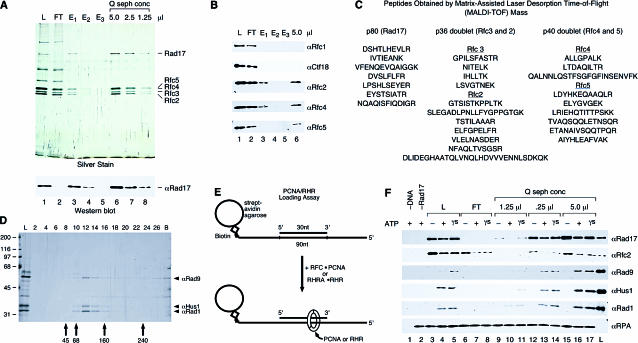
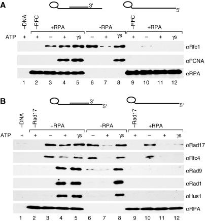
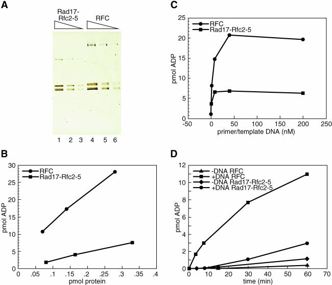
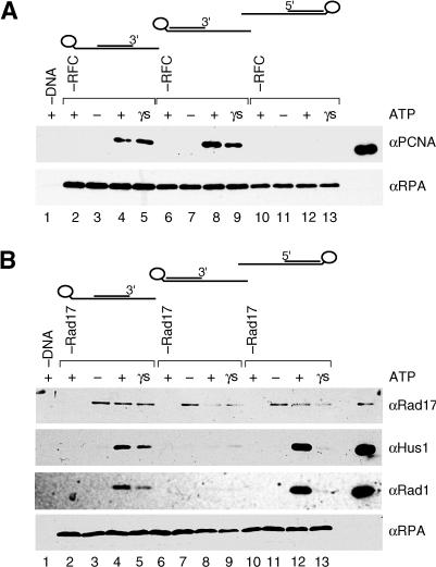
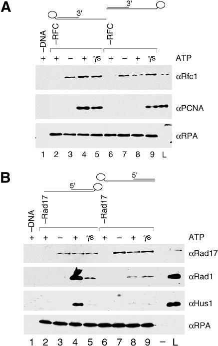
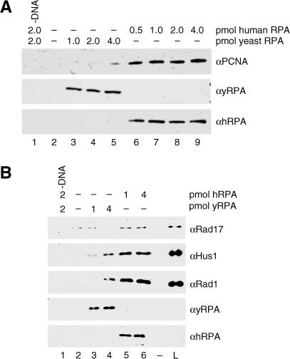
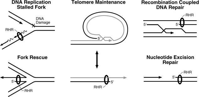
The cellular pathways involved in maintaining genome stability halt cell cycle progression in the presence of DNA damage or incomplete replication. Proteins required for this pathway include Rad17, Rad9, Hus1, Rad1, and Rfc-2, Rfc-3, Rfc-4, and Rfc-5. The heteropentamer replication factor C (RFC) loads during DNA replication the homotrimer proliferating cell nuclear antigen (PCNA) polymerase clamp onto DNA. Sequence similarities suggest the biochemical functions of an RSR (Rad17-Rfc2-Rfc3-Rfc4-Rfc5) complex and an RHR heterotrimer (Rad1-Hus1-Rad9) may be similar to that of RFC and PCNA, respectively. RSR purified from human cells loads RHR onto DNA in an ATP-, replication protein A-, and DNA structure-dependent manner. Interestingly, RSR and RFC differed in their ATPase activities and displayed distinct DNA substrate specificities. RSR preferred DNA substrates possessing 5' recessed ends whereas RFC preferred 3' recessed end DNA substrates. Characterization of the biochemical loading reaction executed by the checkpoint clamp loader RSR suggests new insights into the mechanisms underlying recognition of damage-induced DNA structures and signaling to cell cycle controls. The observation that RSR loads its clamp onto a 5' recessed end supports a potential role for RHR and RSR in diverse DNA metabolism, such as stalled DNA replication forks, recombination-linked DNA repair, and telomere maintenance, among other processes.


Conflict of interest statement

The authors have declared that no conflicts of interest exist.

Figures

Figure 1
Figure 1. The Purified Human RSR Complex Can Load the RHR Complex onto DNA In Vitro
(A) RSR was purified from the Rfc2 Ab affinity column eluate by anti-Rad17 Ab affinity chromatography, and the peptide-eluted material was concentrated by Q–sepharose chromatography. An equivalent volume (5 μl) of the load onto the anti-Rad17 column (lane 1, labeled L), the flowthrough from the column (lane 2, labeled FT), each peptide elution fraction (lanes 3–5), and the indicated amounts of the concentrated, purified complex (lanes 6–8) were analyzed by silver staining and Wb for Rad17. (B) The same fractions present in the silver-stained gel in (A) were analyzed by Wb for Rfc1, Ctf18, Rfc2, Rfc4, and Rfc5, and the lanes are as loaded and numbered in (A). (C) Peptide sequences for the proteins present in the purified Q–sepharose fraction. (D) Purification of the RHR. RHR was purified from E. coli by Talon affinity, Q–sepharose, and phosphocellulose chromatography and by glycerol gradient sedimentation (shown here). The load onto the gradient (lane L) and fractions (corresponding to lane numbers) as well as any material in the pellet (lane B) were analyzed by silver staining (shown) and Wb (not shown). Arrows indicate the sedimentation position of protein standards from a gradient prepared in parallel. (E) Assay for RHR and PCNA loading. RHR and PCNA loading were examined by monitoring the binding of the proteins to a DNA–RPA complex bound to streptavidin–agarose beads by Wb of the bead-bound fractions. The DNA substrate consist of a 90 nucleotide (nt) 3′ biotinylated template and 30 nt primer positioned in the center of the template, resulting in a substrate with 30 nt single-stranded recessed 5′ and 3′ ends to which RPA was bound. (F) RSR is sufficient to load RHR onto DNA in vitro. Reactions were performed as described in the Materials and Methods, and the bead-precipitated products were analyzed by Wb for Rad17, Rfc2, Rad9, Hus1, and Rad1. The fractions from the purification shown in (A) were assayed for RHR-loading activity. Lanes represent reactions that contained the same amount of the anti-Rad17 Ab column load (lanes 1, 3, 4, and 5), flowthrough (lanes 6–8), and the Q–sepharose concentrated protein (lanes 9–17) as shown in the silver-stained gel in (A), or no source of Rad17 (buffer only, lane 2). All reactions contained 5′ and 3′ recessed primer–template DNA–RPA complex bound to beads (except for that in lane 1, which contained beads alone), 1 pmol of RHR complex, and the indicated nucleotide cofactor (ATP: lanes 1, 2, 4, 7, 10, 13, and 16; ATPγS: lanes 5, 8, 11, 14, and 17) or no nucleotide (lanes 3, 6, 9, 12, and 15). The lane labeled L represents 20% of the input of RHR and anti-Rad17 column load used in the reaction.
Figure 3
Figure 3. RHR Loading Is Nucleotide, Primer, and RPA Dependent
Loading reactions represented in (A) were performed with 2 pmol of PCNA, 0.25 pmol of RFC, and the 5′ and 3′ recessed primer–template DNA–RPA complex bound to beads, whereas those in (B) were performed with 1 pmol of RHR, 0.25 pmol of RSR, and the 5′ and 3′ recessed primer–template DNA–RPA complex bound to beads. Reactions were performed as described in the Materials and Methods, and the bead-precipitated proteins were then analyzed by Wb for PCNA, RFC, Rad17, Rad9, Hus1, and Rad1, respectively. In both (A) and (B), lanes 2 and 9 represent reactions that contained the clamp alone (PCNA in [A], RHR in [B]), and all reactions represented in lanes 1, 3–8, and 10–12, contained both the clamp and its corresponding clamp loader. In both (A) and (B), reactions were performed in the absence of nucleotide (lanes 3, 6, and 9) or in the presence of ATP (lanes 1, 2, 4, 7, 9, and 11) or ATPγS (lanes 5, 8, and 12). All reactions contained RPA except those in lanes 6–8, and all reactions contained primer–template DNA bound beads except that in lane 1 (beads without DNA) and those in lanes 6–9 (template DNA alone bound beads).
Figure 2
Figure 2. Purified RSR Is an ATPase That Is Poorly Stimulated by DNA
(A) Titration of purified RSR and RFC. Visualized by SDS-PAGE and silver staining were 0.3, 0.15, and 0.075 pmol of RSR (lanes 1–3) and RFC (lanes 4–6). (B) ATPase activity of the indicated amount of either RSR (squares) or RFC (circles) was measured after 60 min in the presence of 200 nM 5′ and 3′ recessed primer–template DNA. (C) ATPase activity of either 0.3 pmol of RSR (squares) or 0.30 pmol of RFC (circles) was analyzed after a 60 min incubation in the absence or presence of 1.6 nM, 8 nM, 40 nM, or 200 nM 5′ and 3′ recessed primer–template DNA. (D) ATPase activity of 0.15 pmol of RSR (diamonds and circles) or 0.15 pmol of RFC (triangles and squares) in the absence of DNA (diamonds and triangles) or presence of 200 nM 5′ and 3′ recessed primer–template DNA was measured after either a 3.5, 7.5, 15, 30, or 60 min incubation. All reactions were performed as described in the Materials and Methods.
Figure 4
Figure 4. Opposite DNA Substrate Preference Exhibited by RFC and RSR
Loading reactions in (A) and (B) were performed as described in the Materials and Methods with DNA substrates bound to beads, and recovery of proteins with the beads was analyzed by Wb for the indicated proteins. Lanes represent reactions using either a 5′ and 3′ recessed primer–template DNA (lanes 2–5), a 3′ recessed primer–template DNA (lanes 6–9), a 5′ recessed primer–template DNA (lanes 10–13), or no DNA (lane 1). All reactions contained RPA and the indicated nucleotide (ATP: lanes 1, 2, 4, 6, 8, 10, and 12; ATPγS: lanes 5, 9, and 13) or no nucleotide (lanes 3, 7, and 11). In (A), all reactions contained 0.25 pmol of RFC (except for those in lanes 2, 6, and 10) and 2 pmol of PCNA. In (B), all reactions contained 0.25 pmol of RSR (except for those in lanes 2, 6, and 10) and 1 pmol of RHR. In both (A) and (B), 20% of the input in each reaction is represented in the last lane in each panel.
Figure 5
Figure 5. RHR Forms a Sliding Clamp on DNA
Loading reactions in the experiments represented in (A) and (B) were performed as described in the Materials and Methods using either recessed 5′ or recessed 3′ primer–template DNA–RPA substrates bound to beads, and then recovery of proteins with the beads was analyzed by Wb for the indicated proteins. In each experiment using template strands biotinylated at either the 5′ or 3′ end, either both ends of the DNA substrate (reactions represented in lanes 2–5) or only one end of the substrate (reactions represented in lanes 6–9) was blocked by selectively positioning the bead relative to RPA—either at the opposite end of the DNA (distal, therefore both ends blocked) or at the same end of the DNA (proximal, therefore only one end blocked). In (A), lanes represent reactions that contained 0.25 pmol of RFC (except for those in lanes 2 and 6), 2 pmol of PCNA, and either a 3′ recessed primer/3′ biotin template DNA (bead distal to RPA; lanes 2–5), a 3′ recessed primer/5′ biotin template DNA (bead proximal to RPA; lanes 6–9), or no DNA (lane 1). In (B), lanes represent reactions that contained 0.25 pmol of RSR (except for those in lanes 2 and 6), 1 pmol of RHR, and either a 5′ recessed primer/5′ biotin template (bead distal to RPA; lanes 2–5), a 5′ recessed/3′ biotin template (bead proximal to RPA; lanes 6–9) or no DNA (lane 1). In both (A) and (B), reactions were performed in the absence (lanes 3 and 7) or presence of nucleotide (ATP: lanes 2, 4, 6, and 8; ATPγS: lanes 5 and 9), and 20% of the reaction input was loaded in the lane labeled L.
Figure 6
Figure 6. PCNA and RHR Loading Are Specifically Stimulated by Human RPA
In both (A) and (B), lanes represent loading reactions performed as described in the Materials and Methods with either 5′ recessed or 3′ recessed primer–template DNA–RPA complex bound to beads, and recovery of proteins with the beads was analyzed by Wb for the indicated proteins. In (A), PCNA loading was assayed in the absence of RPA (lane 2) or in the presence of the indicated amounts of either yeast RPA (lanes 3–5) or human RPA (lanes 6–9). All reactions contained 3′ recessed primer–template DNA (except for that in lane 1), 2 pmol of PCNA, 0.25 pmol of RFC, and ATP. In (B), RHR loading was assayed in the absence of RPA (lane 2) or in the presence of the indicated amounts of either yeast RPA (lanes 3 and 4) or human RPA (lanes 5 and 6). All reactions contained 5′ recessed primer–template DNA (except for that in lane 1), 1 pmol of RHR complex, 0.25 pmol of RSR, and ATP.
Figure 7
Figure 7. Possible Substrates onto Which the Checkpoint Clamp Loader RSR May Load Its Clamp (RHR)
DNA maintenance pathways, including those depicted here, generate intermediates containing free and/or recessed 3′ ends that are processed by a variety of proteins. These structures also contain recessed 5′ ends, whose fate in these reactions is unclear. Given that RSR loads RHR (depicted as a ring or donut encircling the DNA) onto recessed 5′ ends in vitro, recessed 5′ ends generated in vivo in the depicted pathways can be considered potential substrates. They all contain adjacent single-stranded DNA that could be bound by RPA. RHR has been shown to be required for checkpoint signaling in response to DNA replication fork arrest (Longhese et al. 1997), double-strand breaks (Kondo et al. 2001; Melo et al. 2001), and improper telomere maintenance (Garvik et al. 1995; Lydall and Weinert 1995; Longhese et al. 2000). The RHR clamp is proposed to protect the recessed 5′ end from extensive degradation by exonucleases and to promote resolution of these structures back to duplex DNA.

Similar articles

Cited by

References

    1. Abraham RT. Cell cycle checkpoint signaling through the ATM and ATR kinases. Genes Dev. 2001;15:2177–2196. - PubMed
    1. Bellaoui M, Chang M, Ou J, Xu H, Boone C, et al. Elg1 forms an alternative RFC complex important for DNA replication and genome integrity. EMBO J. 2003;22:4304–4313. - PMC - PubMed
    1. Ben-Aroya S, Koren A, Liefshitz B, Steinlauf R, Kupiec M. ELG1, a yeast gene required for genome stability, forms a complex related to replication factor C. Proc Natl Acad Sci U S A. 2003;100:9906–9911. - PMC - PubMed
    1. Berdis AJ, Benkovic SJ. Mechanism of bacteriophage T4 DNA holoenzyme assembly: The 44/62 protein acts as a molecular motor. Biochemistry. 1997;36:2733–2743. - PubMed
    1. Bermudez VP, Lindsey-Boltz LA, Cesare AJ, Maniwa Y, Griffith JD, et al. Loading of the human 9–1-1 checkpoint complex onto DNA by the checkpoint clamp loader hRad17-replication factor C complex in vitro. Proc Natl Acad Sci U S A. 2003;100:1633–1638. - PMC - PubMed

Publication types

MeSH terms