

Case Reports
Tanycytomas: a newly characterized hypothalamic-suprasellar and ventricular tumor
Affiliations
- PMID: 14625222
- PMCID: PMC8148897
Item in Clipboard
Case Reports
Tanycytomas: a newly characterized hypothalamic-suprasellar and ventricular tumor
AJNR Am J Neuroradiol.
2003 Nov-Dec.
Abstract
We report five cases of tumors occurring in three children and in two adults. The tumors had unusual histomorphology and a mixture of ependymal and piloid-like astrocytic features and a myxoid stroma similar to myxopapillary ependymomas. MR imaging in three of the cases showed aggressive, intensely enhancing partially cystic hypothalamic-suprasellar masses near midline and near the floor of the third ventricle. In the three pediatric cases, the tumor encased the circle of Willis. This newly characterized tumor, the tanycytoma, has neoplastic cells with histomorphologic and ultrastructural characteristics similar to those of the tanycyte.
Figures

Case 1. This 2.5-year-old female patient was unresponsive. A and B, Axial T1-weighted contrast-enhanced images (350/12/2 [TR/TE/NEX]) show a necrotic enhancing suprasellar mass encasing the distal internal carotid arteries. C, Composite image. D, Maximum intensity projection image from 3D time-of-flight MR angiogram, No blood flow is visualized beyond the supraclinoid portions of internal carotid arteries.

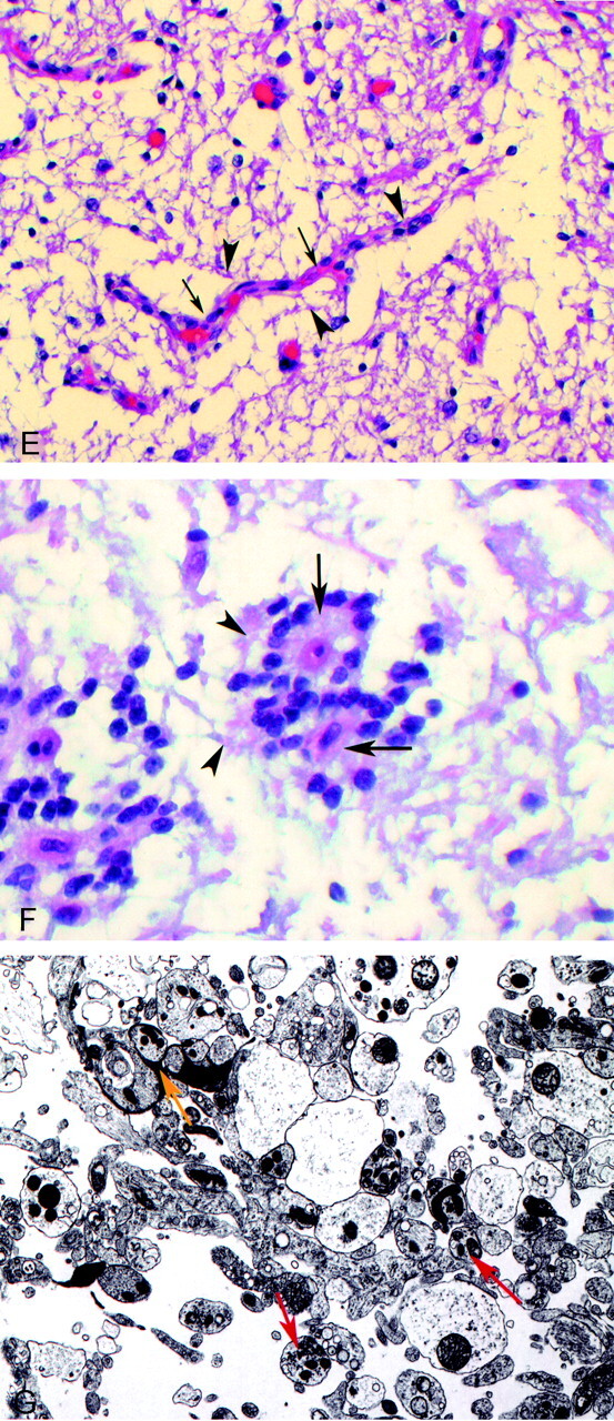
Case 2. Two-year-old male patient with diplopia and a decrease in left eye vision. A, Axial T2-weighted image (3200/84/1) shows a hyperintense suprasellar mass encasing the anterior portion of the circle of Willis. B, Axial T1-weighted image (433/16/2) shows a hypointense suprasellar mass encasing the circle of Willis. C, Contrast-enhanced sagittal T1-weighted image (650/14/2) shows a solid-cystic enhancing hypothalamic-suprasellar mass. D, Contrast-enhanced coronal view T1-weighted image (400/16/2). E, Photomicrograph of hematoxylin and eosin-stained normal tanycytes (arrowheads) radiating to a blood vessel (arrows) in area postrema. F, Photomicrograph of hematoxylin and eosin-stained neoplastic cells (arrowheads) radiating to blood vessel (arrows) in tanycytoma specimen. G, Photomicrograph of electron microscopy of tumor specimen reveals tanycytes with long synaptic processes (red arrow) and synaptoid complexes (yellow arrow).
Case 2. Two-year-old male patient with diplopia and a decrease in left eye vision. A, Axial T2-weighted image (3200/84/1) shows a hyperintense suprasellar mass encasing the anterior portion of the circle of Willis. B, Axial T1-weighted image (433/16/2) shows a hypointense suprasellar mass encasing the circle of Willis. C, Contrast-enhanced sagittal T1-weighted image (650/14/2) shows a solid-cystic enhancing hypothalamic-suprasellar mass. D, Contrast-enhanced coronal view T1-weighted image (400/16/2). E, Photomicrograph of hematoxylin and eosin-stained normal tanycytes (arrowheads) radiating to a blood vessel (arrows) in area postrema. F, Photomicrograph of hematoxylin and eosin-stained neoplastic cells (arrowheads) radiating to blood vessel (arrows) in tanycytoma specimen. G, Photomicrograph of electron microscopy of tumor specimen reveals tanycytes with long synaptic processes (red arrow) and synaptoid complexes (yellow arrow).

Case 4. This 26-year-old female patient presented with galactorrhea and hypothyroidism. A, Sagittal view T1-weighted image (433/8/2) shows a hypointense hypothalamic mass. B, Axial view T2-weighted image (3200/88/2) shows a hyperintense hypothalamic mass with suprasellar extension. C, Coronal view T1-weighted contrast-enhanced image (400/9/2) shows a homogeneously enhancing hypothalamic-suprasellar mass. D, Photomicrograph of tumor specimen stained with glial fibrillary acidic protein shows positive brown-stained glial processes (arrowheads) radiating to blood vessel (arrows). E, Photomicrograph of tumor specimen stained with synaptophysin shows positive brown-stained glial processes (arrowheads) radiating to blood vessel (arrows).
Comment in
-
Suprasellar monomorphous pilomyxoid gliomas.AJNR Am J Neuroradiol. 2003 Nov-Dec;24(10):1931-2. AJNR Am J Neuroradiol. 2003. PMID: 14625211 Free PMC article. No abstract available.
References
-
- Tihan T, Fisher PG, Kepner JL, et al. Pediatric astrocytomas with monomorphous pilomyxoid features and a less favorable outcome. J Neuropathol Exp Neurol 1999;58:1061–1068 - PubMed
-
- Fuller CE, Frankel B, Smith M, et al. Suprasellar monomorphous pilomyxoid neoplasm: an ultrastructural analysis. Clin Neuropathol 2001;6:256–262 - PubMed
-
- Millhouse OE, Zellforsch Z, Mikrosk U. Anat Embryol (Berl) 1971;121:1–13 - PubMed
-
- Mathew TC, Singh DN. Morphology and distribution of tanycytes in the third ventricle of the adult rat: a study using semithin methacrylate sections. Acta Anat (Basel) 1989;134:319–321 - PubMed
-
- Maruyama R, Koga K, Nakahara T, Kishida K, Nabeshima K. Cerebral myxopapillary ependymoma. Hum Pathol 1992;23:960–962 - PubMed
Publication types
MeSH terms
LinkOut - more resources
Full Text Sources
Medical