

Case Reports
Imaging of acute bilateral paramedian thalamic and mesencephalic infarcts
Affiliations
- PMID: 14625223
- PMCID: PMC8148919
Item in Clipboard
Case Reports
Imaging of acute bilateral paramedian thalamic and mesencephalic infarcts
AJNR Am J Neuroradiol.
2003 Nov-Dec.
Abstract
Thalami and midbrain arterial supply arises from many perforating blood vessels with a complex distribution for which many variations have been described. One rare variation, named the "artery of Percheron," is a solitary arterial trunk that arises from one of the proximal segments of a posterior cerebral artery and supplies the paramedian thalami and the rostral midbrain bilaterally. Occlusion of this artery results in bilateral thalamic and mesencephalic infarctions. We describe three patients with a presumed occlusion of the artery of Percheron in whom MR imaging showed characteristic symmetrical bilateral paramedian thalamic and mesencephalic infarctions.
Figures
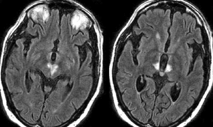
Case 1. Axial FLAIR images (8500/110/2500/1 [TR/TE/TI/NEX]) show infarcts in the medial inferior thalami and extending into the medial and superior midbrain (territory of the artery of Percheron).
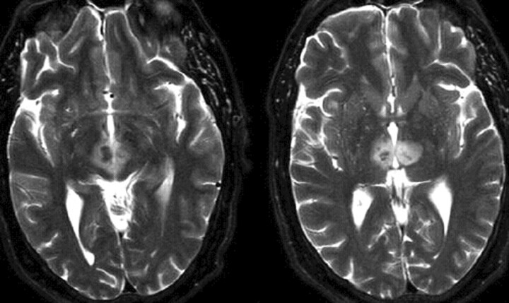
Case 2. Axial T2-weighted images (5800/99/1 [TR/TE/NEX]) show areas of increased signal intensity in the paramedian thalamic and midbrain regions. Within the infarcts, there are hypointense areas suggesting the presence deoxyhemoglobin secondary to hemorrhages.
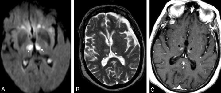
Case 3. A, Axial trace diffusion-weighted image (5700/138 [TR/TE], b = 1000 s/mm2) obtained 24 hours after the onset of symptoms shows bilateral thalamic areas of high signal intensity (white arrows) compatible with that of acute paramedian thalamic infarcts. B, Axial T2-weighted image (5800/99/1 [TR/TE/NEX]) shows rounded areas (arrowheads) of increased signal intensity in the medial thalami. C, Axial T1-weighted (440/17/1 [TR/TE/NEX]) postcontrast image obtained 12 days after image in panel A shows contrast enhancement in the bilateral thalamic infarcts (black arrowheads).
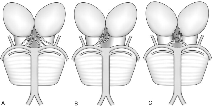
Variations of the paramedian thalamic-mesencephalic arterial supply according to Percheron. A, In the most common variation, there are many small perforating arteries arising from the P1 segments of the PCA. B, The artery of Percheron is a single perforating blood vessel arising from one P1 segment. C, The third type of variation is that of an arcade of perforating branches arising from an artery bridging the P1 segments of both PCAs.
References
-
- Percheron G. The anatomy of the arterial supply of the human thalamus and its use for the interpretation of the thalamic vascular pathology. Z Neurol 1973;205:1–13 - PubMed
-
- Lasjaunias P, Berenstein A, Brugge KGT, eds. Surgical Neuroangiography. 2nd ed. Berlin: Springer-Verlag;2000. ,Vol. 1:526–562
-
- Roitberg BZ, Tuccar E, Alp MS. Bilateral paramedian thalamic infarct in the presence of an unpaired thalamic perforating artery. Acta Neurochir 2002;144:301–304 - PubMed
-
- Lepore FD, Gulli V, Miller DC. Neuro-ophthalmological findings with neuropathological correlation in bilateral thalamic-mesencephalic infarction. J Clin Neuro-ophthalmol 1985;5:224–228 - PubMed
-
- Kumral E, Evyapan D, Balkir K, et al. Bilateral thalamic infarction, clinical etiological and MRI correlates. Acta Neurol Scand 2001;103:35–42 - PubMed
Publication types
MeSH terms
LinkOut - more resources
Full Text Sources
Medical