

A novel image fusion visualizes the angioarchitecture of the perforating arteries in the brain
Affiliations
- PMID: 14625225
- PMCID: PMC8148933
Item in Clipboard
A novel image fusion visualizes the angioarchitecture of the perforating arteries in the brain
AJNR Am J Neuroradiol.
2003 Nov-Dec.
Abstract
We report a novel technique that fuses 3D digital subtraction angiograms and MR images. Image fusion was successfully performed within 20 minutes each in 11 consecutive cases. Our initial experience showed that this image fusion enabled clear and simultaneous visualization of perforating arteries and surrounding tissues. The relation between perforating arteries and normal brain or lesions was easily understood in a clinical setting by using this image fusion.
Figures
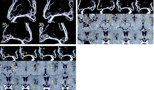
Images in a patient with no pathology (contralateral side of a right ICA aneurysm, which was treated with endovascular obliteration). A, 3D DSA (volume rendering) image, which can be seen stereoscopically, clearly shows one left RAH and one left LSA (upper, anteroposterior view; lower, posteroanterior view). B, Common cursor image, which simultaneously displays 3D DSA (MIP), axial, and coronal contrast-enhanced MR images, clearly shows the passing course of the left RAH (short arrow, origin of the RAH; long arrow, top of the cranial loop; arrowhead, immediately before entering the anterior perforating substance; double arrow, location at the putamen). The corresponding points are indicated as small red dots. C, Common cursor image clearly shows the passing course of the left LSA (short arrow, location in the sylvian fissure; long arrow, location at the anterior perforating substance; arrowhead and double arrow, locations at the putamen). The corresponding points are indicated as small red dots.
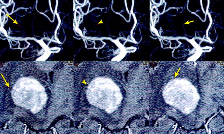
Common cursor image in a patient with a right thalamic malignant lymphoma. The image, which simultaneously displays 3D DSA (MIP) and axial contrast-enhanced MR images, clearly shows the relation between the tumor and two LSAs (long arrow and arrowhead) or one RAH (short arrow). The corresponding points are indicated as small red dots.
Similar articles
-
Role of image fusion combining three-dimensional digital subtraction angiography with magnetic resonance imaging in evaluation of unruptured cerebral aneurysms.Neurol Res. 2007 Jan;29(1):58-63. doi: 10.1179/174313206X153806. Neurol Res. 2007. PMID: 17427277
-
Usefulness of 3D DSA-MR fusion imaging in the pretreatment evaluation of brain arteriovenous malformations.Acad Radiol. 2012 Nov;19(11):1345-52. doi: 10.1016/j.acra.2012.07.001. Epub 2012 Aug 28. Acad Radiol. 2012. PMID: 22951112
-
Visualization of the intracisternal angioarchitecture at the posterior fossa by use of image fusion.Neurosurgery. 2005 Feb;56(2):335-42; discussion 335-42. doi: 10.1227/01.neu.0000148005.29708.1c. Neurosurgery. 2005. PMID: 15670381
-
[Advances in the contributions of imaging to stereotaxic localization of cerebral arteriovenous malformations for radiosurgery ].Neurochirurgie. 2001 May;47(2-3 Pt 2):201-11. Neurochirurgie. 2001. PMID: 11404697 Review. French.
-
[Advanced vascular imaging techniques of supra-aortic, encephalic and medullary vessels].J Radiol. 2007 Mar;88(3 Pt 2):472-82. doi: 10.1016/s0221-0363(07)89847-7. J Radiol. 2007. PMID: 17457258 Review. French.
Cited by
-
Perforating arteries originating from the posterior communicating artery: a 7.0-Tesla MRI study.Eur Radiol. 2009 Dec;19(12):2986-92. doi: 10.1007/s00330-009-1485-4. Eur Radiol. 2009. PMID: 19533146 Free PMC article.
-
Optimization of the Surgical Approach in AVMs Using MRI and 4D DSA Fusion Technique : A technical note.Clin Neuroradiol. 2017 Dec;27(4):443-450. doi: 10.1007/s00062-017-0571-2. Epub 2017 Mar 13. Clin Neuroradiol. 2017. PMID: 28289757
-
Evaluation of CT angiography for visualisation of the lenticulostriate artery: difference between normotensive and hypertensive patients.Br J Radiol. 2012 Nov;85(1019):e1004-8. doi: 10.1259/bjr/67294268. Epub 2012 Jun 27. Br J Radiol. 2012. PMID: 22744324 Free PMC article.
-
Diagnostic accuracy of three-dimensional-rotational angiography and heavily T2-weighted volumetric magnetic resonance fusion imaging for the diagnosis of spinal arteriovenous shunts.J Neurointerv Surg. 2022 Jan;14(1):neurintsurg-2020-017252. doi: 10.1136/neurintsurg-2020-017252. Epub 2021 Mar 4. J Neurointerv Surg. 2022. PMID: 33674393 Free PMC article.
-
Analyzing characteristics of collateral flow to parasylvian cortical arteries by three-dimensional digital subtraction angiography-magnetic resonance angiography fusion imaging in adult moyamoya disease.Front Neurol. 2023 Sep 21;14:1251844. doi: 10.3389/fneur.2023.1251844. eCollection 2023. Front Neurol. 2023. PMID: 37808502 Free PMC article.
References
-
- Anxionnat R, Bracard S, Ducrocq X, et al. Intracranial aneurysms: clinical value of 3D digital subtraction angiography in the therapeutic decision and endovascular treatment. Radiology 2001;218:799–808 - PubMed
-
- Marinkovic S, Gibo H, Milisavljevic M, Cetkovic M. Anatomic and clinical correlations of the lenticulostriate arteries. Clin Anat 2001;14:190–195 - PubMed
-
- Gorczyca W, Mohr G. Microvascular anatomy of Heubner’s recurrent artery. Neurol Res 1987;9:259–264 - PubMed
MeSH terms
LinkOut - more resources
Full Text Sources
Medical