

Review
Calcification of the olfactory bulbs in three patients with hyposmia
Affiliations
- PMID: 14625241
- PMCID: PMC8148898
Item in Clipboard
Review
Calcification of the olfactory bulbs in three patients with hyposmia
AJNR Am J Neuroradiol.
2003 Nov-Dec.
Abstract
An estimated two million Americans suffer from chemosensory disorders. We present the clinical and imaging findings in three hyposmic patients with bilateral olfactory bulb calcification detected by CT. To our knowledge, these are the first cases of olfactory bulb calcification reported in the literature. A review of the literature concerning calcification of cranial nerves, olfactory neuritis, and the potential etiology and clinical significance of olfactory bulb calcification in our patients is presented.
Figures
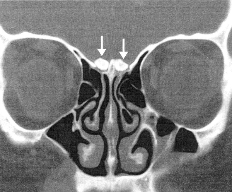
Coronal CT scan from patient 1 demonstrates attenuated, rounded calcifications above the cribriform plate (arrows) in the location of the olfactory bulbs.
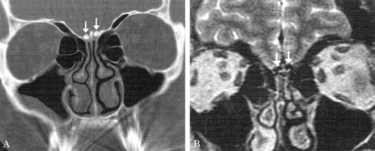
Patient 2. A, Coronal CT image shows attenuated calcification in the location of the olfactory bulbs (arrows) similar to that seen in patient 1. B, Coronal T2-weighted MR image demonstrates markedly hypointense signal in the olfactory bulbs (arrows) corresponding to the areas of calcification noted on the corresponding CT scan (A). No other anterior cranial fossa lesions were identified.
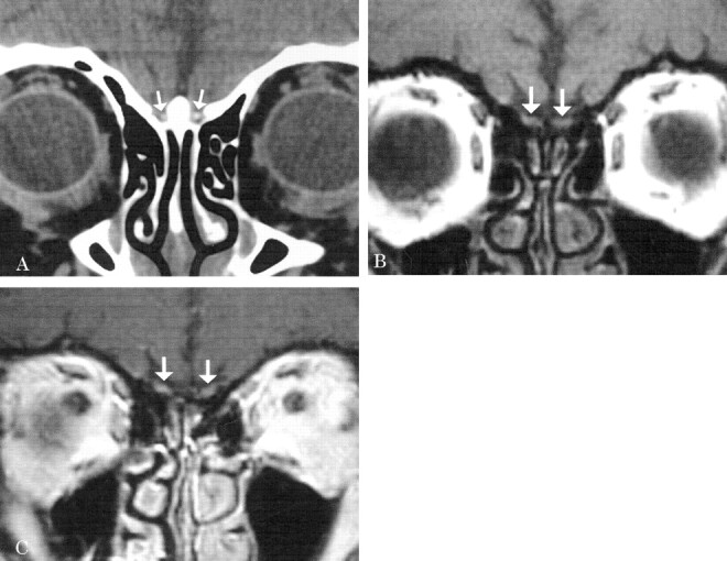
Patient 3. A, Coronal CT scan shows punctate calcifications in the expected location of the olfactory bulbs (arrows). B, Coronal T1-weighted MR image demonstrates the olfactory bulbs (arrows) before contrast agent administration. C, Postcontrast coronal T1-weighted MR image demonstrates mild enhancement of both olfactory bulbs (arrows).
Similar articles
-
Posttraumatic smell loss: relationship of psychophysical tests and volumes of the olfactory bulbs and tracts and the temporal lobes.Acad Radiol. 1999 May;6(5):264-72. doi: 10.1016/s1076-6332(99)80449-8. Acad Radiol. 1999. PMID: 10228615
-
Magnetic resonance imaging of the olfactory apparatus.Arch Otolaryngol Head Neck Surg. 1994 Aug;120(8):869-72. doi: 10.1001/archotol.1994.01880320069015. Arch Otolaryngol Head Neck Surg. 1994. PMID: 8049051
-
Systemic lupus erythematosus associated with marked intracranial calcification.AJNR Am J Neuroradiol. 1992 Sep-Oct;13(5):1340-2. AJNR Am J Neuroradiol. 1992. PMID: 1414826 Free PMC article.
-
Olfactory bulb volume in the clinical assessment of olfactory dysfunction.Rhinology. 2009 Mar;47(1):3-9. Rhinology. 2009. PMID: 19382487 Review.
-
Imaging of chemosensory loss.Otolaryngol Clin North Am. 2004 Dec;37(6):1255-80, vii. doi: 10.1016/j.otc.2004.06.008. Otolaryngol Clin North Am. 2004. PMID: 15563913 Review.
Cited by
-
Olfaction disorders: retrospective study.Braz J Otorhinolaryngol. 2014 Jan-Feb;80(1):11-7. doi: 10.5935/1808-8694.20140005. Braz J Otorhinolaryngol. 2014. PMID: 24626886 Free PMC article. Review. English, Portuguese.
References
-
- Schiffman SS. Taste and smell in disease. N Engl J Med 1983;308:1337–1343 - PubMed
-
- Seiden AM. The initial assessment of patients with taste and smell disorders. In: Seiden AM, ed. Taste and Smell Disorders. 1st ed. New York: Thieme;1997. :4–19
-
- Duncan HJ, Smith DV. Clinical disorders of olfaction: a review. In: Doty RL, ed. Handbook of Olfaction and Gustation. 1st ed. New York: Marcel Dekker;1995. :345–365
-
- Truwit CL, Kelly WM. The olfactory system. Neuroimaging Clin North Am 1993;3:47–70
-
- Jafek BW, Hartman D, Eller PM, et al. Postviral olfactory dysfunction. Am J Rhinol 1990;4:91–100
Publication types
MeSH terms
LinkOut - more resources
Full Text Sources
Medical