Laparoscopic suture repair of a perforated gastric ulcer in a severely cirrhotic patient with portal hypertension: first case report
- PMID: 14626407
- PMCID: PMC3021346
Laparoscopic suture repair of a perforated gastric ulcer in a severely cirrhotic patient with portal hypertension: first case report
Abstract
Background: Open digestive surgery in cirrhotic patients is associated with high morbidity and mortality. Laparoscopy in this setting has the potential to reduce postoperative complications. Laparoscopic treatment of a perforated gastric ulcer in a severely cirrhotic patient with portal hypertension is herein described.
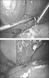
Methods: A 75-year-old woman affected by cirrhosis of the liver (Child class C) and chronic gastric ulcer presented with acute abdominal pain. The diagnosis of perforation was made with plain films of the abdomen and computed tomography. Diagnostic laparoscopy showed intense peritonitis due to a perforated ulcer of the anterior gastric wall, 2 cm proximal to the pylorus. Suture closure and placement of an omental patch were performed laparoscopically.
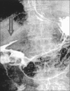
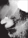
Results: Postoperative recovery was complicated by a minor leak of the gastric suture, managed by total parenteral nutrition. Closure of the gastric wound was demonstrated by Gastrografin studies on the 10th postoperative day. The patient was discharged on the 16th postoperative day. At 3-months follow-up, the patient is alive and free of gastric disease.
Figures


Similar articles
-
Transumbilical single-access laparoscopic perforated gastric ulcer repair.Surg Innov. 2012 Jun;19(2):130-3. doi: 10.1177/1553350611423931. Epub 2011 Oct 23. Surg Innov. 2012. PMID: 22025424
-
Laparoscopic repair of perforated gastroduodenal ulcer by running suture.Langenbecks Arch Surg. 2004 Feb;389(1):11-6. doi: 10.1007/s00423-003-0427-x. Epub 2003 Nov 15. Langenbecks Arch Surg. 2004. PMID: 14618328
-
[Laparoscopic closure and tegmentation of perforated ulcer].Tidsskr Nor Laegeforen. 1992 May 20;112(13):1696-7. Tidsskr Nor Laegeforen. 1992. PMID: 1387266 Norwegian.
-
Perforated gastric remnant ulcer after laparoscopic gastric bypass.BMJ Case Rep. 2018 Apr 5;2018:bcr2017222190. doi: 10.1136/bcr-2017-222190. BMJ Case Rep. 2018. PMID: 29622702 Free PMC article. Review.
-
Do perforated duodenal ulcers need an acid-decreasing surgical procedure now that omeprazole is available?Surg Clin North Am. 1992 Apr;72(2):369-80. doi: 10.1016/s0039-6109(16)45684-7. Surg Clin North Am. 1992. PMID: 1549799 Review.
Cited by
-
Nanofibrillar cellulose-alginate hydrogel coated surgical sutures as cell-carrier systems.PLoS One. 2017 Aug 22;12(8):e0183487. doi: 10.1371/journal.pone.0183487. eCollection 2017. PLoS One. 2017. PMID: 28829830 Free PMC article.
-
Laparoscopic correction of perforated peptic ulcer: first choice? A review of literature.Surg Endosc. 2010 Jun;24(6):1231-9. doi: 10.1007/s00464-009-0765-z. Epub 2009 Dec 24. Surg Endosc. 2010. PMID: 20033725 Free PMC article. Review.
-
Outcomes of abdominal surgery in patients with liver cirrhosis.World J Gastroenterol. 2016 Mar 7;22(9):2657-67. doi: 10.3748/wjg.v22.i9.2657. World J Gastroenterol. 2016. PMID: 26973406 Free PMC article. Review.
References
-
- Schwartz SI. Biliary tract surgery and cirrhosis. A critical combination. Surgery. 1981;90:577–83 - PubMed
-
- Sleeman D, Namias N, Levi D, et al. Laparoscopic cholecystectomy in cirrhotic patients. J Am Coll Surg. 1998;187(4):400–403 - PubMed
-
- Lacy AM, Balaguer C, Andrade E, et al. Laparoscopic cholecystectomy in cirrhotic patients. Surg Endosc. 1995;9:407–408 - PubMed
-
- Aranha GV, Sontag SJ, Greenlee HB. Cholecystectomy in cirrhotic patients: a formidable operation. Am J Surg. 1982; 143:55–60 - PubMed
Publication types
MeSH terms
LinkOut - more resources
Full Text Sources
Medical
