. 2003 Summer;8(2):134-43.
doi: 10.1379/1466-1268(2003)008<0134:apctpd>2.0.co;2.

Antigenic peptides complexed to phylogenically diverse Hsp70s induce differential immune responses

Affiliations

Antigenic peptides complexed to phylogenically diverse Hsp70s induce differential immune responses

Udayasankar Kumaraguru et al. Cell Stress Chaperones. 2003 Summer.

Abstract

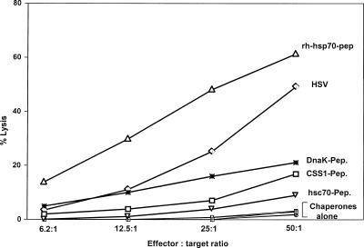
The Hsp70 class of heat shock proteins (Hsps) has been implicated at multiple points in the immune response, including initiation of proinflammatory cytokine production, antigen recognition and processing, and phenotypic maturation of antigen-presenting cells (APCs). This class of chaperones is highly conserved in both sequence and structure, from prokaryotes to higher eukaryotes. In all cases, these chaperones function to bind short segments of either peptides or proteins through an adenosine triphosphate-dependent process. In addition to a possible role in antigen presentation, these chaperones have also been proposed to function as a potent adjuvant. We compared 4 evolutionary diverse Hsp70s, E. coli DnaK, wheat cytosolic Hsc70, plant chloroplastic CCS1, and human Hsp70, for their ability to prime and augment a primary immune response against herpes simplex virus-1 (HSV1). We discovered that all 4 Hsp70s were highly effective as adjuvants displaying similar ability to lipopolysaccharides in upregulating cytokine gene expression. In addition, they were all capable of inducing phenotypic maturation of APCs, as measured by the display of various costimulatory molecules. However, only the human Hsp70 was able to mediate sufficient cross-priming activity to afford a protective immune response to HSV1, as judged by protection from a lethal viral challenge, in vitro proliferation, cytotoxicity, and intracellular interferon-gamma production. The difference in immune response generated by the various Hsp70s could possibly be due to their differential ability to interact productively with other coreceptors and different regulatory cochaperones.


Figures

Fig 1.
Fig 1.
Zoster challenge of mice immunized with SSIEFARL-loaded chaperones. Five-week-old to 6-week-old C57BL/6 mice were scarified on their left flank after depilating with a hair clipper and a chemical depilator (Nair-Carter, Wallace, NY, USA). The mice were anesthetized using metofane (Pitman-Moore, Mundelein, IL, USA), and a total of 20 scarifications were made in an approximately 4-mm2 area. To such scarifications, 10 μL of PBS containing 106 plaque-forming units of herpes simplex virus-1 (HSV1) strain 17 was added and gently massaged. Animals were inspected daily for the development of zosteriform ipsilateral lesions, general behavioral changes, encephalitis, and mortality. The severity of the lesions was scored as follows: 1+ = vesicle formation; 2+ = local erosion and ulceration of the local lesion; 3+ = mild to moderate ulceration; 4+ = severe ulceration, hind limb paralysis, and encephalitis; 5 = moribund animals that were euthanized. Numbers in parentheses denote average scores. The figure shows individual scores for 10 mice in each group.
Fig 2.
Fig 2.
(a) Staining for activation marker. Splenocytes (106) were stained with labeled anti-CD80, anti-CD86, and anti-CD40 after stimulation in vitro with various heat shock proteins (Hsps). Unstimulated splenocytes were stained with the same antibodies to serve as background for histogram overlay. The cells were washed and analyzed using FACScan and CellQuest software. The highest amount of endotoxin found in the Hsp preparations (1 EU) was used as a control to show the contribution of lipopolysaccharide (LPS) contamination to upregulation of activation markers. Controls included were Hsps that were individually either heat inactivated or protease treated (data not shown). The figure represents 1 of 5 experiments with similar patterns of results. (b) Cytokine analysis by enzyme-linked immunosorbent assay (ELISA). The supernatants collected from adherent splenocyte cultures stimulated with various Hsps were collected and analyzed by ELISA. The concentrations of each cytokine were deduced from the standard curve obtained using recombinant cytokine. One EU of LPS, the contaminating amount found in one of the preparations, was used as control for background. Supernatants collected from unstimulated, heat-inactivated, and protease-treated cultures served as controls (data not shown). The figure represents 1 of 5 experiments with similar patterns of results
Fig 3.
Fig 3.
Hsp70 obtained from various species loaded in vitro with SSIEFARL also induces HSVgB498–505 SSIEFARL-specific CD8+ T cell proliferative response in mice. Five-week-old to 6-week-old C57BL/6 mice were immunized with SSIEFARL-loaded recombinant human Hsp70, DnaK, Hsc70, and CSS1, and SSIEFARL-conjugated bovine serum albumin and HSV1 kos (1 × 10⁶ plaque-forming units) on day 0 and day 21. One week later, the splenocytes were harvested and nylon wool nonadherent T cells were assessed for in vitro proliferative response to SSIEFARL peptide–pulsed antigen-presenting cells (APCs). The responders (2 × 10⁶) were serially double diluted, and the stimulators (1 × 10⁵) were mixed at a ratio starting from 20:1 (responder-stimulator), with the addition of 50 U/mL of recombinant interleukin-2, and incubated for 5 days, with the last 18 hours in the presence of [3H]-thymidine. The cells were harvested and read with an Inotech automatic cell harvester and reader and the results expressed as counts per minute. The controls included and not shown are anti-CD3–stimulated responders, stimulators only, and responders with irrelevant peptide-pulsed APCs. The figure represents the average of 5 mice
Fig 4.
Fig 4.
Immunization of B6 mice with Hsp70-SSIEFARL induces interferon-γ (IFNγ)–positive SSIEFARL-specific CD8+ T cells. Intracellular staining for IFNγ: 1 × 10⁶ splenocytes obtained from immunized C57BL/6 mice were stimulated in vitro with the SSIEFARL peptide (1 μg/mL), with the addition of 50 U of recombinant interleukin-2 and 1 μg/mL of Brefeldin A for 6 hours. The cells were later washed and stained for CD8 with phycoerythrin-labeled anti-CD8 and then washed again, fixed, permeabilized, and stained intracellularly for IFNγ with fluorescein isothiocyanate–labeled anti-IFNγ. The controls included isotype antibody control. The stained cells were analyzed with a FACScan machine. The CD8+ IFNγ+ T cells are seen in the upper right quadrant. The experiment was repeated 3 times, which gave similar results. The figure represents one such experiment
Fig 5.
Fig 5.
In vivo immunization with Hsp70-peptide induces potent primary cytotoxic T lymphocyte (CTL) responses. B6 mice were immunized with SSIEFARL-loaded recombinant human Hsp70, DnaK, Hsc70, and CSS1; SSIEFARL-conjugated bovine serum albumin and HSV1 kos (1 × 10⁶ plaque-forming units); or buffer only intraperitoneally on day 0, boosted on day 21, and terminated on day 28 or day 60 after the second immunization for spleen cell collection. A portion of the single-cell suspension of the spleen was in vitro expanded for 5 days at 37°C and 5% CO2, with X-ray–irradiated (3000 rads), herpes simplex virus (HSV)–infected (1.5 moi), or SSIEFARL peptide–pulsed (5 μg/mL) B6 antigen-presenting cells. At the end of the incubation period, they were used as effectors in a chromium release CTL assay. 51Cr-pulsed, major histocompatibility complex (MHC)–matched, HSV-infected (MC38-HSV); MHC-matched, SSIEFARL-pulsed (MC38-SSIEFARL); MHC-mismatched, HSV-infected, and SSIEFARL-pulsed (EMT-6-HSV, EMT6-SSIEFARL); and MHC-matched, uninfected (MC38) targets were used. The figure shows the percentage of lysis for MC38-SSIEFARL targets only and the results of 3 different experiments

Similar articles

Cited by

References

    1. Arnold D, Faath S, Rammensee H, Schild H. Cross-priming of minor histocompatibility antigen-specific cytotoxic T cells upon immunization with the heat shock protein gp96. J Exp Med. 1995;182:885–889. - PMC - PubMed
    1. Asea A, Kraeft SK, Kurt-Jones EA, Stevenson MA, Chen LB, Finberg RW, Koo GC, Calderwood SK. HSP70 stimulates cytokine production through a CD14-dependant pathway, demonstrating its dual role as a chaperone and cytokine. Nat Med. 2000;6:435–442. - PubMed
    1. Basu S, Binder RJ, Ramalingam T, Srivastava PK. CD91 is a common receptor for heat shock proteins gp96, hsp90, hsp70, and calreticulin. Immunity. 2001;14:303–313. - PubMed
    1. Basu S, Binder RJ, Suto R, Anderson KM, Srivastava PK. Necrotic but not apoptotic cell death releases heat shock proteins, which deliver a partial maturation signal to dendritic cells and activate the NF-kappa B pathway. Int Immunol. 2000;12:1539–1546. - PubMed
    1. Beech JT, Siew LK, Ghoraishian M, Stasiuk LM, Elson CJ, Thompson SJ. CD4+ Th2 cells specific for mycobacterial 65-kilodalton heat shock protein protect against pristane-induced arthritis. J Immunol. 1997;159:3692–3697. - PubMed

Publication types

LinkOut - more resources