Case Reports
. 2003 Nov 19;23(33):10548-58.
doi: 10.1523/JNEUROSCI.23-33-10548.2003.

Pathogenesis of X-linked Charcot-Marie-Tooth disease: differential effects of two mutations in connexin 32

Affiliations
Case Reports

Pathogenesis of X-linked Charcot-Marie-Tooth disease: differential effects of two mutations in connexin 32

Charles K Abrams et al. J Neurosci. .

Abstract

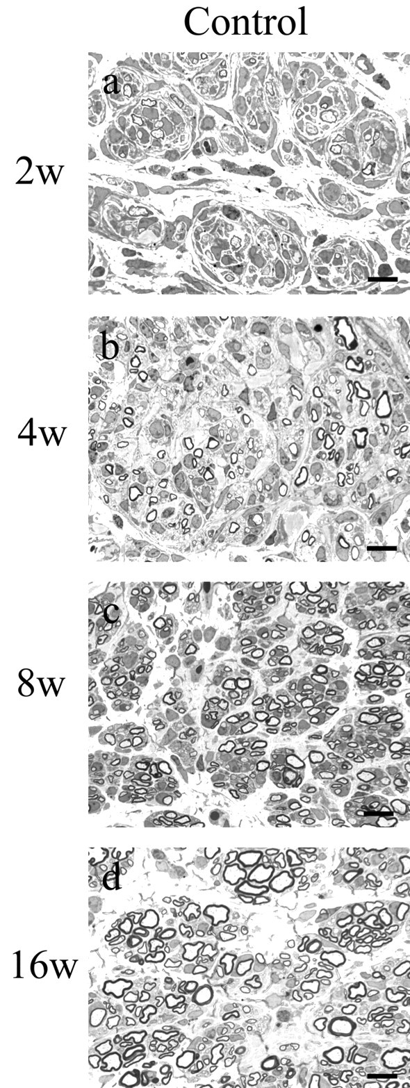
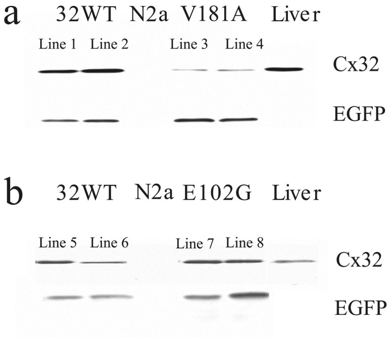
X-linked Charcot-Marie-Tooth disease is an inherited peripheral neuropathy arising in patients with mutations in the gene encoding connexin 32 (Cx32). Cx32 is expressed at the paranodes and Schmidt-Lantermann incisures of myelinating Schwann cells in which it is believed to form a reflexive pathway between the abaxonal and adaxonal cytoplasmic domains. Patients with the Val181Ala (V181A) mutation have a severe peripheral neuropathy. Experiments using a nude mouse xenograft system show that Schwann cells expressing only this mutant form of Cx32 are profoundly impaired in their ability to support the earliest stages of regeneration of myelinated fibers. Coupling between paired Xenopus oocytes expressing V181A is reduced compared with the coupling between oocytes expressing wild-type human Cx32 (32WT), and protein levels assayed by Western blot are substantially lower. Immunocytochemisty shows that Neuro2a cells expressing the V181A mutant have very few gap junction plaques compared with cells expressing 32WT; Cx32 protein levels are lower in these cells than in those expressing 32WT. Because failure of normal regeneration is evident before formation of myelin, loss of function of Cx32 may impact on the function of precursors of the myelinating Schwann cell before the formation of the hypothesized reflexive pathway. The Glu102Gly (E102G) mutation leads to a milder phenotype. Early regeneration is normal in grafts with Schwann cells expressing the E102G mutant. The only abnormality detected in the behavior of its channel is increased sensitivity to acidification-induced closure, a property that may lead to reduced gap junction coupling during periods of metabolic stress. This restricted functional abnormality may explain the relatively mild phenotype seen in the xenograft model and in E102G patients.


Figures

Figure 1.
Figure 1.
Sural nerve biopsies from male patients harboring mutations in Cx32. a, Sural nerve biopsy from a patient with the V181A mutation. Prominent features include the presence of thinly myelinated smaller-diameter fibers and regeneration-associated onion bulbs (arrowheads). Scattered enlarged periaxonal spaces (arrows) were seen in this biopsy specimen. The inset shows a fiber associated with an enlarged periaxonal space and onion bulb. b, Sural nerve biopsy from a patient with the E102G mutation. Fibers with normal myelin thickness as well as regeneration-associated onion bulbs (arrowhead) were seen in this biopsy. c, MF size distribution in sural nerve biopsies. Quantitative morphological analyses of the nerves were performed as described in Materials and Methods. The fiber loss was more severe in the V181A nerve with an MF density of 4872/mm2 in comparison with the E102G nerve (6174/mm2) and an age-matched control nerve (8038/mm2). In both mutations, there were larger numbers of small-diameter myelinated axons, often in clusters, suggesting active regeneration, and there were fewer axons >4 μm. The large-diameter fibers in the V181A mutation comprised only 3% of the total MFs in the V181A nerve compared with 11% in the E102G nerve and 60% in the control nerve. Scale bars, 10 μm.
Figure 2.
Figure 2.
Regeneration in control xenografts at 2 (a), 4 (b), 8 (c), and 16 (d) weeks after surgical procedures. One-micrometer-thick, toluidine blue-stained cross-sections taken ∼1 mm proximal to the distal graft host junction are shown. Sural nerve was obtained from the normal donor, and nerve xenografts were generated as described in Materials and Methods. By 2 weeks, many myelinated axons, predominantly in the 2-4 μm size range, had regenerated into the graft. The density of all fiber sizes increased at 4 and 8 weeks, but a relative increase in the number of very small (<2 μm) MFs was noted. At 16 weeks, there was a substantial increase in the number of large (>4 μm)-diameter fibers (for histograms, see Fig.4a-d). Scale bars, 10 μm.
Figure 3.
Figure 3.
Regeneration in V181A and E02G xenografts at 2, 4, 8, and 16 weeks after surgical procedures. One-micrometer-thick, toluidine blue-stained cross-sections taken ∼1 mm proximal to the distal graft host junction are shown. a-d, V181A xenografts at 2 (a), 4 (b), 8 (c), and 16 (d) weeks. Regeneration was significantly delayed compared with the E102G and control xenografts. Occasional fibers showed regions of increased periaxonal space (c, d, arrowheads). e-h, E102G xenografts at 2 (e), 4 (f), 8 (g), and 16 (h) weeks. Axons efficiently regenerated into and through the xenograft. Enlarged regions of periaxonal space noted for the V181A xenografts were not present in the E102G grafts. Although more prominent in the more proximal portion of the graft (Sahenk and Chen, 1998), abnormally enlarged axons were also seen in the distal segments of the E102G grafts. These were most prominent at 8 weeks. Scale bars, 10 μm.
Figure 4.
Figure 4.
MF size distribution in xenografts. Quantitative morphological analysis of xenografts at 2, 4, 8, and 16 weeks. a-d, MF size distribution histograms at 4, 8, and 16 weeks after grafting revealed significantly different patterns of regeneration-associated myelination for these two Cx32 mutations. At 2 and 4 weeks, regeneration and myelination of all fiber sizes through the V181A grafts were significantly retarded compared with E102G and controls grafts. At 8 and 16 weeks, V181A densities approached those for E102G but remained below those in control grafts. e, Total MF densities for control and E102G were similar at 2 and 4 weeks. At later times, the densities for control, but not E102G, MFs continued to increase. MF counts for V181A xenografts were reduced at all time points.
Figure 5.
Figure 5.
Expression of 32WT and the V181A and E102G mutants in Xenopus oocytes. a, Graphic representation of junctional conductance of homotypically paired oocytes injected with antisense oligonucleotide to Cx38 (Anti38) or mRNA for 32WT, or the V181A or E102G mutants determined as described in Materials and Methods; n = 44, 26, 37, and 15, respectively. b, Representative Western blot of total protein extracts from sister oocytes of those injected for assessment of junctional conductance. Levels of expression of V181A were significantly lower than those of 32WT (see Results for details).
Figure 6.
Figure 6.
Representative current traces and average normalized Gj-Vj relationships for 32WT and the E102G and V181A mutants. a, c, e, Xenopus oocytes were injected with the noted mRNAs and paired as described in Materials and Methods. Junctional currents recorded from cell 1 in response to voltage steps to cell 2 in 20 mV increments from ±20 to ±120 are shown. b, d, f, Average instantaneous (▵) and steady-state (▪) Gj-Vj relationships for 32WT and the two mutants determined from currents like those in a, c, and e and plotted as mean ± SEM. g-i, Average normalized Gj-Vj relationships for 32WT and the two mutants paired heterotypically with 26WT. Xenopus oocytes were injected with the noted mRNAs and paired heterotypically with 26WT. Junctional currents in response to a series of voltage steps were obtained and analyzed. The resulting instantaneous (▵) and steady-state (▪) Gj-Vj relationships were averaged and plotted as mean ± SEM.
Figure 7.
Figure 7.
Single-channel recordings of the cell-cell channel formed by the V181A and E102G mutants. Ramps were applied to cell 1 from -120 to +120 mV, and currents were recorded in cell 2. a, Single-channel currents from cells expressing the V181A mutant. b, Single-channel currents from cells expressing the E102G mutant. c, Graphic representation of the voltage waveform applied to cell 1. The sizes of the major transitions (arrows) for both channels (∼50-55 pS) were similar to what we (data not shown) and others (Oh et al., 1997) have observed for 32WT.
Figure 8.
Figure 8.
Dye transfer between Neuro2a cells transfected with the V181A*EGFP or E102G*EGFP mutants. Pairs of cells expressing fluorescent plaques, a morphological correlate of EGFP-tagged gap junctions, were selected. Using the dual patch-clamp technique, high-resistance seals were established with each of the adjacent cells expressing V181A*EGFP (a) and E102G*EGFP (d). One pipette (patched to cell 1, denoted by an asterisk) contained both recording solution and ∼1 mm Lucifer yellow, whereas the other pipette, patched to cell 2, contained only recording solution. The patch under the Lucifer yellow-containing pipette was then ruptured, allowing dye to diffuse into cell 1. b, e, Fluorescent images obtained a few seconds after rupture. c,f, Fluorescent images obtained ∼30 sec later for V181A and 90 sec later for E102G, showing substantial amounts of Lucifer yellow in cell 2 of each pair. At the end of each experiment, the membrane under pipette 2 was ruptured, and junctional conductance was measured (in this case, 50 nS for the V181A-expressing pair and 25 nS for the E102G-expressing pair). In addition, the presence of a cytoplasmic bridge was excluded by demonstrating that the conductance between cell 1 and cell 2 could be reduced to undetectable levels by application of a bath solution containing 1% heptanol (v/v). Similar findings were obtained with 32WT*EGFP. In no case did untransfected Neuro2a cells lacking cytoplasmic bridges show detectable levels of dye transfer.
Figure 9.
Figure 9.
Effect of bath acidification with CO2 on junctional conductance of 32WT and the V181A and E102G mutants. Oocytes were injected with the mRNAs for 32WT, V181A, and E102G and paired homotypically. Both cells were clamped at -30 mV, and junctional conductances were monitored by measuring the current responses in cell 2 to a ±10 mV pulse applied to cell 1 (frequency, 0.5 Hz). After establishing a stable baseline conductance for at least 2 min, the bath was acidified by continuous perfusion with bath solution saturated with CO2 (arrows). Twelve minutes later, the perfusate was changed to a standard bath solution at pH 7.6 (arrowheads). Junctional conductances were determined at 30 sec intervals and plotted for times beginning 1 min before application of CO2-saturated solution. As shown, the effects of acidification and realkalinization on the 32WT-induced (a) and V181A-induced (b) junctional conductances were nearly identical, whereas the E102G-induced (c) junctional conductance declines more rapidly and fully; the E102G-induced conductance also shows a longer delay in onset of recovery when CO2 is removed from the bath. The overshoot seen with realkalinization of the WT and E102G was quite variable; overshoot was not as prominent with the V181A mutant, in which overall protein levels are lower. Overshoot is likely an effect of pH on junctional conductance distinct from any direct effect on channel gating.
Figure 10.
Figure 10.
Expression levels of 32WT and E102G in transfected Neuro2a cells were significantly higher than that of V181A expressed in stably transfected Neuro2A cells, as determined by Western blot. Neuro2A cells were stably transfected with 32WT or the V181A or E102G mutants in the pIRES-EGFP vector as described in Materials and Methods. Protein extracts from 5 × 10⁴ cells/well were loaded in each lane, and blots were probed with Cx32 antibody as described in Materials and Methods. a, Comparison of two cell lines expressing Cx32WT (1 and 2) or the V181A mutation (3 and 4). As shown, levels of EGFP expression in the four lines are comparable. However, the level of expression of V181A is much lower than that of 32WT. b, Comparison of two lines expressing Cx32WT (5 and 6) or the E102G mutation (7 and 8). As shown, levels of expression of EGFP in the two lines are comparable as are levels of 32WT and E102G.
Figure 11.
Figure 11.
Immunohistochemical localization of Cx32 in stably transfected Neuro2a cells expressing 32WT or the E102G or V181A mutants. a, 32WT is distributed in intercellular and intracellular puncta as well as, too a lesser extent, diffusely in the cytoplasm in a predominantly perinuclear distribution. The intercellular puncta are likely to be gap junction plaques. b, Like 32WT, the E102G mutant shows large intercellular plaques; interestingly, the number of intracellular puncta seems reduced. c, V181A is distributed diffusely, predominantly in the perinuclear cytoplasm with extremely rare intracellular or intercellular puncta. d, These control cells expressing Cx32WT were processed in a manner identical to those shown in a-c, except that mouse IgG was substituted for the Cx32-specific antibody. Similar results were obtained using Cx32-specific antibody with untransfected Neuro2a cells.

Similar articles

Cited by

References

    1. Abrams CK, Bennett MVL ( 2000) Hereditary human diseases caused by connexin mutations. In: Gap junctions—molecular basis of cell communication in health and disease (Peracchia C, ed) pp 423-459. New York: Academic.
    1. Abrams CK, Freidin MM, Dobrenis K, Bargiello TA, Verselis VK, Bennett MVL, Sahenk Z ( 2000) Pathogenesis of CMTX: analysis of a new connexin 32 mutation leading to an inability to regenerate large caliber axons. Ann Neurol 48: 439.
    1. Abrams CK, Freidin MM, Verselis VK, Bennett MVL, Bargiello TA ( 2001) Functional alterations in gap junction channels formed by mutant forms of connexin 32: evidence for loss of function as a pathogenic mechanism in the X-linked form of Charcot-Marie-Tooth disease. Brain Res 900: 9-25. - PMC - PubMed
    1. Altevogt BM, Kleopa KA, Postma FR, Scherer SS, Paul DL ( 2002) Connexin 29 is uniquely distributed within myelinating glial cells of the central and peripheral nervous systems. J Neurosci 22: 6458-6470. - PMC - PubMed
    1. Anzini P, Neuberg DH, Schachner M, Nelles E, Willecke K, Zielasek J, Toyka KV, Suter U, Martini R ( 1997) Structural abnormalities and deficient maintenance of peripheral nerve myelin in mice lacking the gap junction protein connexin 32. J Neurosci 17: 4545-4551. - PMC - PubMed

Publication types

MeSH terms