. 2003 Nov 19;23(33):10622-32.
doi: 10.1523/JNEUROSCI.23-33-10622.2003.

Activation of erbB-1 signaling in tanycytes of the median eminence stimulates transforming growth factor beta1 release via prostaglandin E2 production and induces cell plasticity

Affiliations

Activation of erbB-1 signaling in tanycytes of the median eminence stimulates transforming growth factor beta1 release via prostaglandin E2 production and induces cell plasticity

Vincent Prevot et al. J Neurosci. .

Abstract

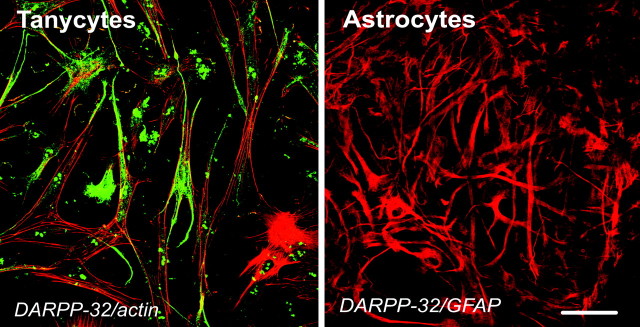
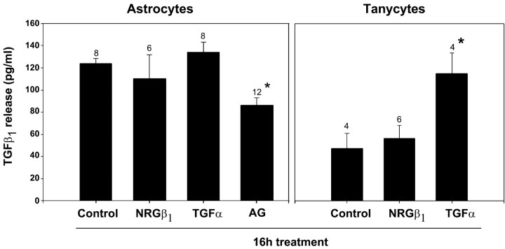
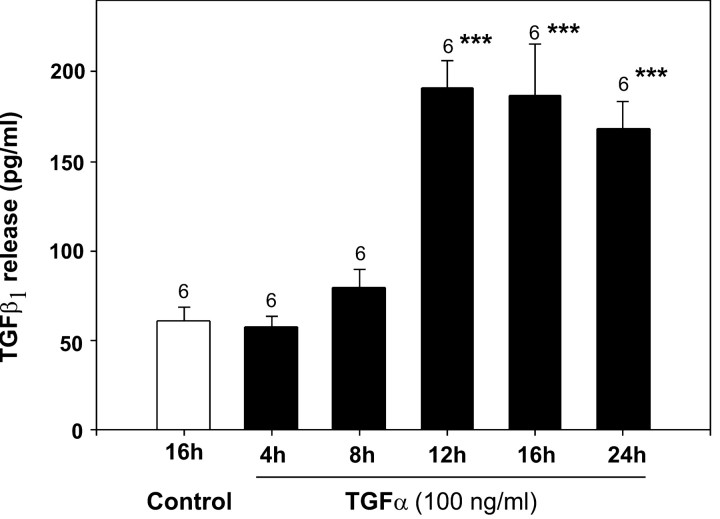
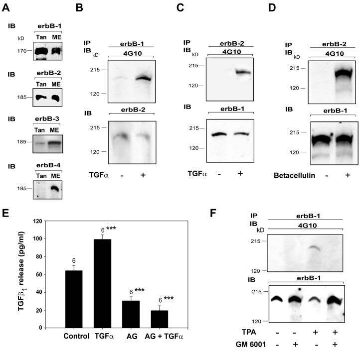
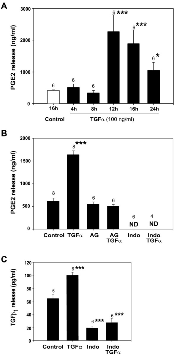
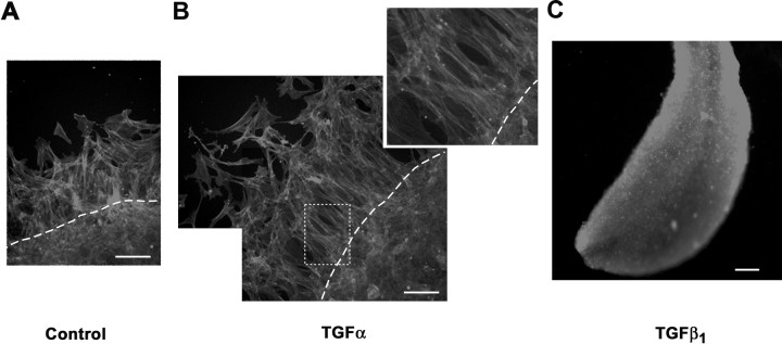
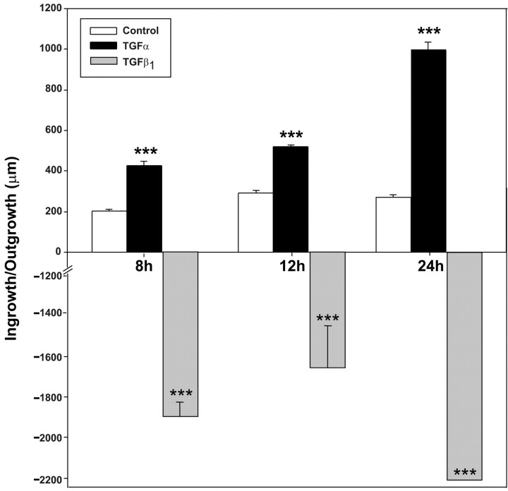
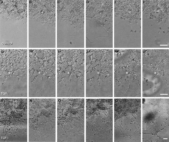
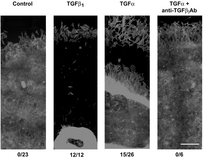
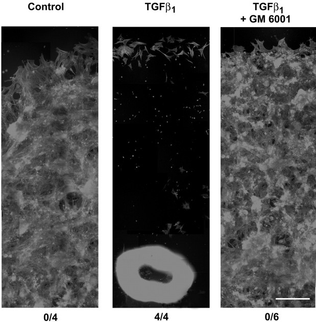
The activation of transforming growth factor alpha (TGFalpha)-erbB-1 and neuregulin-erbB-4 signaling pathways in hypothalamic astrocytes has been shown to play a key role in the process by which the neuroendocrine brain controls luteinizing hormone-releasing hormone (LHRH) secretion. Earlier studies suggested that tanycytes, an ependymoglial cell type of the median eminence, regulate LHRH release during the estrous cycle by undergoing plastic changes that alternatively allow or prevent direct access of the LHRH nerve terminals to the portal vasculature. Neither the molecules responsible for these plastic changes nor the underlying controlling mechanisms have been identified. Here we show that cultured tanycytes express erbB-1 and erbB-2, two of the four members of the erbB receptor family, and respond to TGFalpha with receptor phosphorylation, release of prostaglandin E2 (PGE2), and a PGE2-dependent increase in the release of TGFbeta1, a growth factor previously implicated in the glial control of LHRH secretion. Blockade of either erbB-1 receptor signal transduction or prostaglandin synthesis prevented the stimulatory effect of TGFalpha on both PGE2 and TGFbeta1 release. Time-lapse studies revealed that TGFalpha and TGFbeta1 have dramatically opposite effects on tanycyte plasticity. Whereas TGFalpha promotes tanycytic outgrowth, TGFbeta1 elicits retraction of tanycytic processes. Blockade of metalloproteinase activity abolished the effect of TGFbeta1, suggesting that TGFbeta1 induces tanycytic retraction by facilitating dissolution of the extracellular matrix. Prolonged (>12 hr) exposure of tanycytes to TGFalpha resulted in focal tanycytic retraction, an effect that was abolished by immunoneutralization of TGFbeta1 action, indicating that the retraction was attributable to TGFalpha-induced TGFbeta1 formation. These in vitro results identify tanycytes as targets of TGFalpha action and demonstrate that activation of erbB-1-mediated signaling in these cells results in plastic changes that, involving PGE2 and TGFbeta1 as downstream effectors, mimic the morphological plasticity displayed by tanycytes during the hours encompassing the preovulatory surge of LHRH.


Figures

Figure 1.
Figure 1.
DARPP-32 immunostaining in primary cultures of tanycytes and astrocytes, the two major glial cell types present in the median eminence of the hypothalamus. Although high levels of immunoreactive DARPP-32 are detected in tanycytes cultured in ADM (left panel, green), DARPP-32-immunoreactivity is absent in GFAP-positive cultures of hypothalamic astrocytes (right panel, red). F-actin was visualized in tanycytes (left panel, red) by exposing the fixed and permeabilized cells to phalloidin (diluted 1:150) for 20 min at room temperature. Scale bar, 10 μm.
Figure 2.
Figure 2.
TGFα (100 ng/ml) stimulates TGFβ1 release from tanycytes (left panel) but not astrocytes (right panel) cultured in ADM for 16 hr as assessed by ELISA. AG1478 (left panel, AG, 50 μm), an inhibitor of erbB-1 receptor-tyrosine kinase activity, significantly reduces TGFβ1 production by hypothalamic astrocytes, indicating that activation of erbB-1 signaling is required for basal TGFβ1 release in these glial cells. NRGβ1, another member of the EGF family, is ineffective in stimulating TGFβ1 secretion from either hypothalamic astrocytes or tanycytes. *p < 0.05 versus basal release (control). In this and subsequent figures, columns are means; error bars indicate SEM; and the numbers on top of the vertical lines are the numbers of independent observations per group.
Figure 3.
Figure 3.
Time course of the stimulatory effect of TGFα (100 ng/ml) on TGFβ1 release from cultured tanycytes. ***p < 0.001 versus control.
Figure 4.
Figure 4.
Tanycytes (Tan) of the median eminence (ME) contain erbB-1, erbB-2, and low levels of erbB-3 but not erbB-4 receptors (A). For erbB-1 and erbB-2 detection, each lane was loaded with 25 μg of protein; for erbB-3 detection, the tanycyte lane was loaded with 50 μg of protein, and the ME lane was loaded with 100 μg. Activation of erbB-1 signaling with either TGFα (B, C) or betacellulin (D) results in tyrosine phosphorylation of the erbB-1-erbB-2 heterodimeric complex. Tanycytes were cultured in defined medium and were exposed to erbB-1 ligands (100 ng/ml) for 5 min. Proteins were collected after treatment and immunoprecipitated (IP) with specific erbB-1 (B) or erbB-2 (C, D) antibodies, electrophoresed to size fractionate the immunoprecipitated species, and immunoblotted (IB) with antibodies to phosphotyrosine (4G10). Then immunoblots were stripped and reprobed with antibodies to erbB-2 (B) or erbB-1 (C, D). Note that immunoprecipitation of erbB-1 receptors coimmunoprecipitates erbB-2 receptors (B), and, conversely, immunoprecipitation of erbB-2 receptors coimmunoprecipitates erbB-1 (C, D), even in the absence of ligand stimulation. E, Exposure of tanycytic cultures to AG1478 (50 μm), an inhibitor of erbB-1 signaling, significantly blocks both basal TGFβ1 production (control) and TGFα-induced TGFβ1 release (***p < 0.001 vs control). F, Activation of PKC with TPA results in the metalloproteinase-dependent transphophorylation of erbB-1 receptors in tanycytes of the median eminence. Tanycytes were treated for 5 min with TPA (1 μm). To block metalloproteinase activity, some cultures were pretreated for 30 min with GM6001 (50 μm). After treatment, erbB-1 receptors were immunoprecipitated, and the phosphorylated species were detected by immunoblotting with antibody 4G10. Then the immunoblot was stripped and reprobed with antibodies to erbB-1 to ensure that equal amounts of erbB-1 protein had been immunoprecipitated and loaded on the gel.
Figure 5.
Figure 5.
TGFα-induced TGFβ1 secretion in tanycytes of the median eminence requires PGE2 release. A, TGFα (100 ng/ml) increases PGE2 production in tanycytes cultured in ADM after 12 hr of treatment as assessed by RIA. B, TGFα-induced PGE2 release is blocked by the addition of AG1478 (50 μm), an erbB-1-tyrosine kinase blocker, to the culture medium and abolished by the addition of indomethacin (50 μm), an inhibitor of cyclooxygenase activity. C, Indomethacin obliterates both basal and TGFα-induced TGFβ1 secretion from tanycytes (50 μm). *p < 0.05; ***p < 0.001 versus control.
Figure 6.
Figure 6.
Ability of TGFα and TGFβ1 to induce plastic rearrangements of isolated tanycytes in culture as assessed by monolayer migration assays. Under basal unstimulated conditions, circular monolayers of tanycytes cultured in ADM for 24 hr exhibit a definite but limited ability to move from preset locations in the culture well (dotted lines) toward more peripheral locations (A). TGFα (50 ng/ml, 24 hr) dramatically increases the ability of tanycytes to extend processes and to migrate (B). Note that tanycytic processes exhibited bundles of actin filaments (red) arranged in parallel along the entire longitudinal axis of the process (B, inset). Activation of TGFβ signaling with TGFβ1 (0.4 nm) elicits a complete retraction of tanycytes within 12 hr (C). In all panels the actin cytoskeleton of tanycytes was labeled using Texas Red-labeled phalloidin and was visualized by fluorescent microscopy. Scale bars, 200 μm.
Figure 7.
Figure 7.
Quantitative morphometric analysis of the plastic changes elicited by TGFα and TGFβ1 on circular monolayers of tanycytes fixed after 8, 12, or 24 hr of treatment. ***p < 0.001 versus control.
Figure 8.
Figure 8.
Time-lapse recording of TGFα and TGFβ1 effects on tanycytic morphology in circular cell monolayers perifused with Leibovitz's L15 medium for 12 hr. Under basal unstimulated conditions, tanycytes exhibit motility, but the overall movement of the cells at the edge of the circular monolayer is very limited (A-F). Treatment of tanycytes with TGFα (50 ng/ml) strongly stimulates cell motility and induces pronounced cell shape remodeling (G-L). Arrowheads in G-L point to a tanycytic end foot specialization that undergoes filopodial formation (H-J, arrows) and process extension (K, L, arrows) during the treatment period. When tanycytic cultures are exposed to TGFβ1 (0.4 nm), circular cell monolayers undergo dramatic retraction (M-R). In P, arrows point to tanycytes whose processes stretch between their initial adhesion point on the laminin-coated glass coverslip and the new inward location of the monolayer. These processes eventually retract in Q (arrows). Scale bars, 200 μm.
Figure 9.
Figure 9.
Exposure of circular monolayers of tanycytes to TGFα for 24 hr results in a TGFβ1-dependent retraction of the cells in 60% of the cultures. Photo montages of representative cultures illustrate these findings. Under basal unstimulated conditions (control), no retraction of the circular monolayer was observed in 23 cultures examined (0/23). TGFα (50 ng/ml, 24 hr) and TGFβ1 (0.4 nm, 24 hr) induced retraction in 60% (15/26) and 100% (12/12) of the cultures, respectively. When neutralizing antibodies to TGFβ1 (5 μg/ml) were added for 24 hr to tanycytic cultures treated with TGFα (50 ng/ml), none of the cultures (0/6) showed monolayer retraction. In all panels, the actin cytoskeleton of tanycytes was labeled using Texas Red-labeled phalloidin and was visualized by fluorescent microscopy. Scale bars, 800 μm.
Figure 10.
Figure 10.
Blockade of matrix metalloproteinase activity abolishes the effect of TGFβ1 on tanycyte retraction, as assessed by migration assays. To block matrix metalloproteinase activity, we treated cultures with GM6001 (50 μm). Cotreatment of tanycytic cultures with TGFβ1 (0.4 nm) and GM6001 (50 μm) for 24 hr abrogated the ability of TGFβ1 to promote tanycyte retraction. In all panels, the actin cytoskeleton of tanycytes was labeled using Texas Red-coupled phalloidin and was visualized by fluorescent microscopy. At the bottom of each panel, the left number indicates the number of cultures that retracted under the given experimental condition, and the right number indicates the total number of cultures that were analyzed. Scale bars, 800 μm.

References

    1. Baskin G, Schenker S, Frosto T, Henderson G ( 1981) Transforming growth factor β1 inhibits epidermal growth factor receptor endocytosis and down-regulation in cultured fetal rat hepatocytes. J Biol Chem 266: 13238-13242. - PubMed
    1. Beauvillain JC, Tramu G, Garaud JC ( 1984) Coexistence of substances related to enkephalin and somatostatin in granules of the guinea-pig median eminence: demonstration by use of colloidal gold immunocytochemical methods. Brain Res 301: 389-393. - PubMed
    1. Berens ME, Rief MD, Loo MA, Giese A ( 1994) The role of extracellular matrix in human astrocytoma migration and proliferation studied in a microliter scale assay. Clin Exp Metastasis 12: 405-415. - PubMed
    1. Buchanan CD, Mahesh VB, Brann DW ( 2000) Estrogen-astrocyte-luteinizing hormone-releasing hormone signaling: a role for transforming growth factor-β. Biol Reprod 62: 1710-1721. - PubMed
    1. Chauvet N, Parmentier ML, Alonso G ( 1995) Transected axons of adult hypothalamoneuro-hypophysial neurons. J Neurosci Res 41: 129-144. - PubMed

Publication types

MeSH terms