. 2003 Nov 19;23(33):10672-80.
doi: 10.1523/JNEUROSCI.23-33-10672.2003.

Emerging role for autophagy in the removal of aggresomes in Schwann cells

Affiliations

Emerging role for autophagy in the removal of aggresomes in Schwann cells

Jenny Fortun et al. J Neurosci. .

Abstract

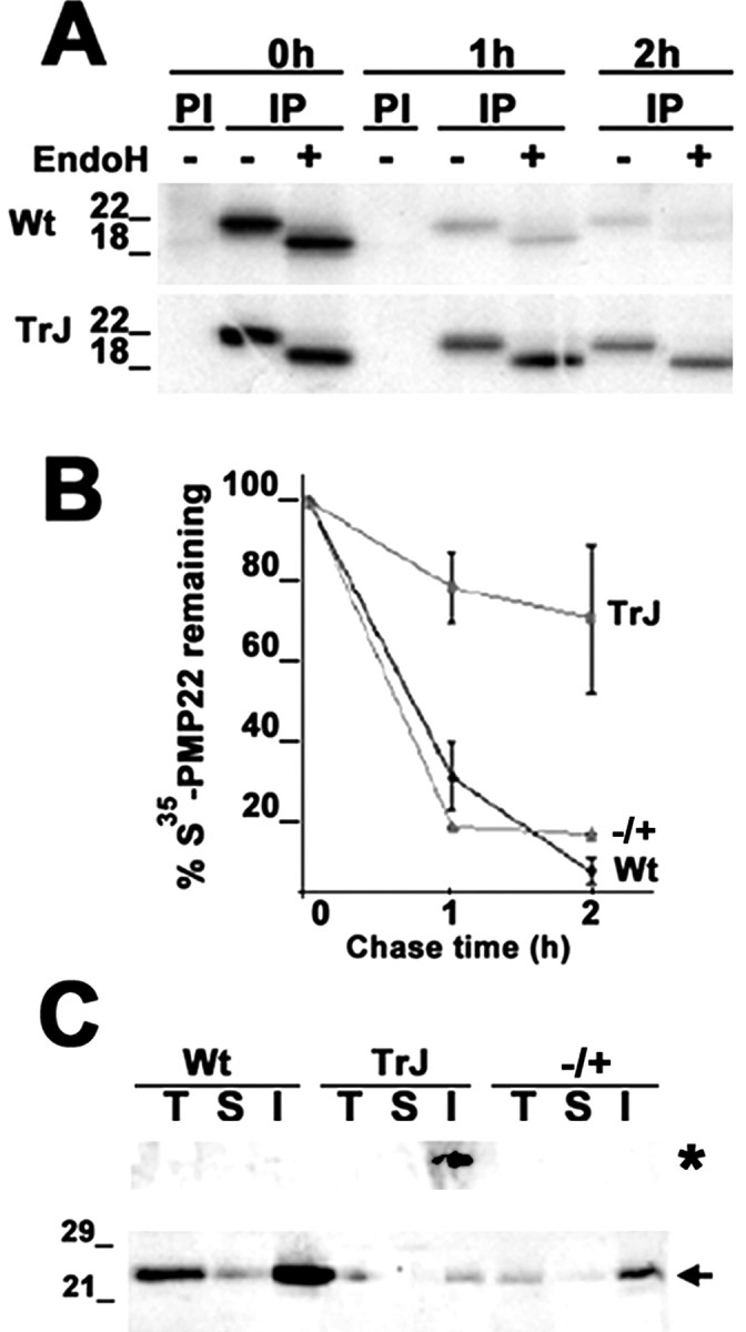
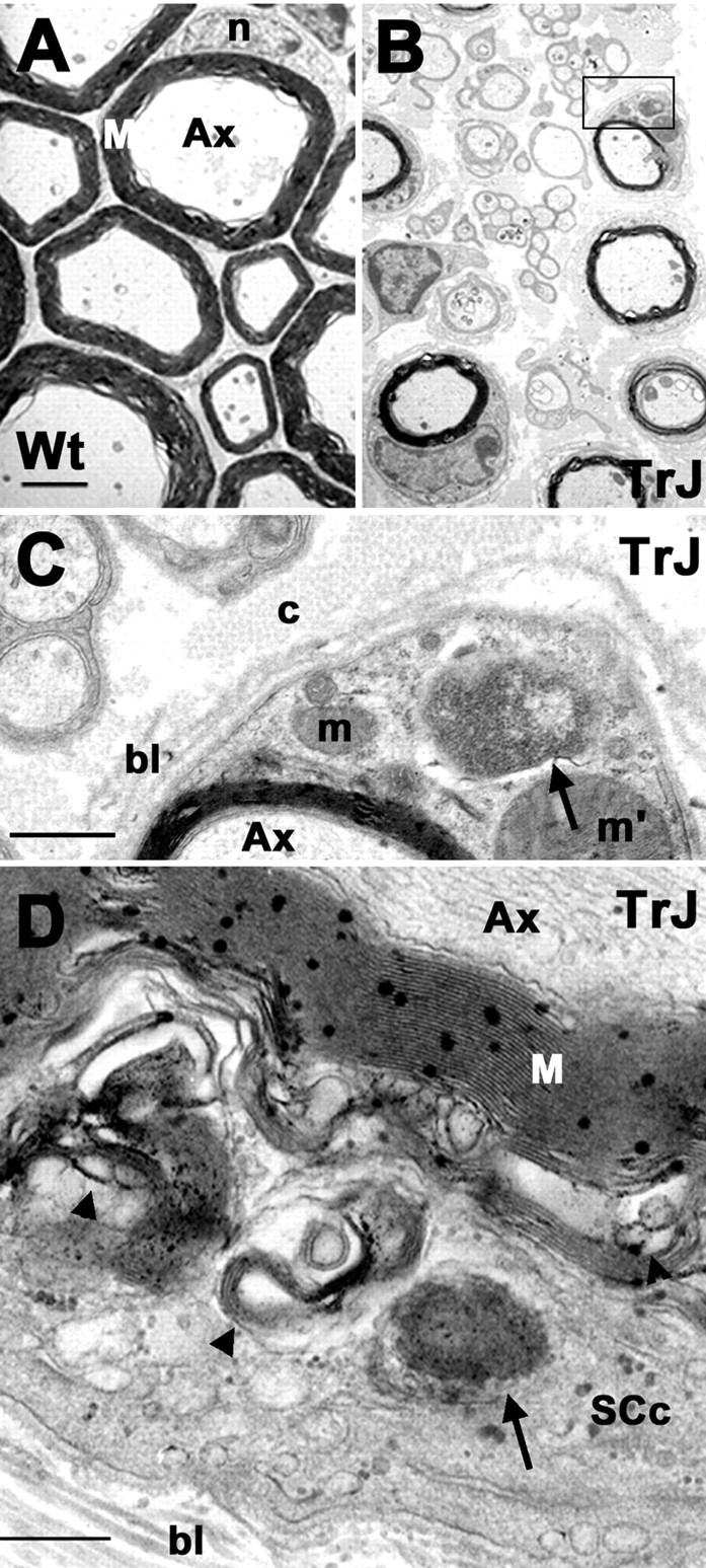
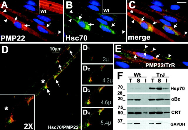
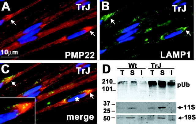
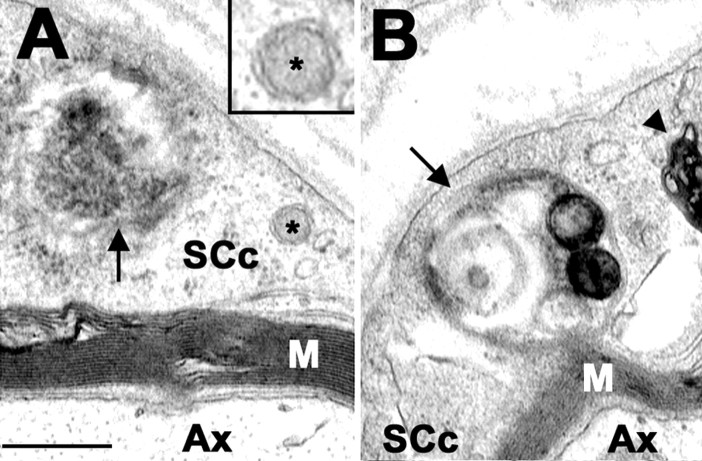
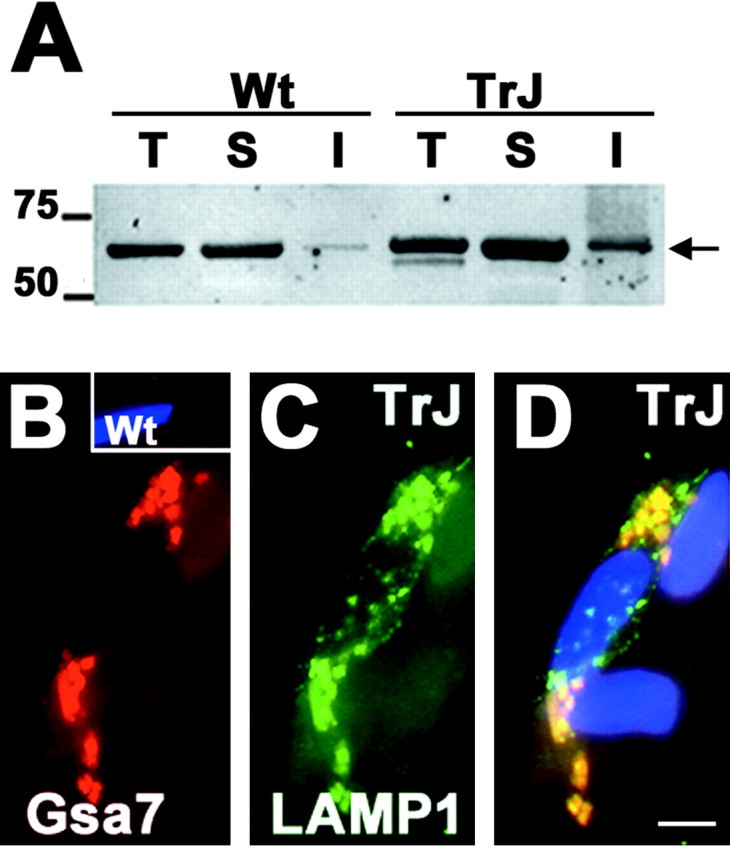
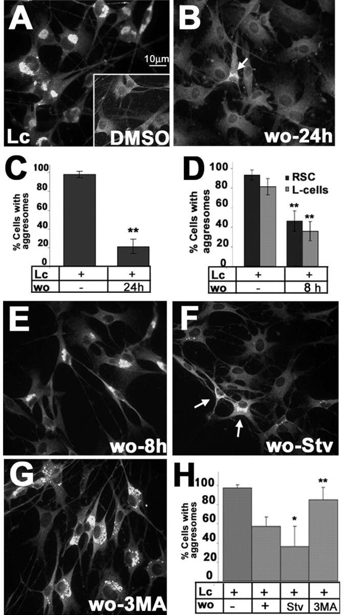
The presence of protein aggregates in the nervous system is associated with various pathological conditions, yet their contribution to disease mechanisms is poorly understood. One type of aggregate, the aggresome, accumulates misfolded proteins destined for degradation by the ubiquitin-proteasome pathway. Peripheral myelin protein 22 (PMP22) is a short-lived Schwann cell (SC) protein that forms aggresomes when the proteasome is inhibited or the protein is overexpressed. Duplication, deletion, or point mutations in PMP22 are associated with a host of demyelinating peripheral neuropathies, suggesting that, for normal SC cell function, the levels of PMP22 must be tightly regulated. Therefore, we speculate that mutant, misfolded PMP22 might overload the proteasome and promote aggresome formation. To test this, sciatic nerves of Trembler J (TrJ) neuropathy mice carrying a leucine-to-proline mutation in PMP22 were studied. In TrJ neuropathy nerves, PMP22 has an extended half-life and forms aggresome-like structures that are surrounded by molecular chaperones and lysosomes. On the basis of these characteristics, we hypothesized that PMP22 aggresomes are transitory, linking the proteasomal and lysosomal protein degradation pathways. Here we show that Schwann cells have the ability to eliminate aggresomes by a mechanism that is enhanced when autophagy is activated and is primarily prevented when autophagy is inhibited. This mechanism of aggresome clearance is not unique to peripheral glia, because L fibroblasts were also capable of removing aggresomes. Our results provide evidence for the involvement of the proteasome pathway in TrJ neuropathy and for the role of autophagy in the clearance of aggresomes.


Figures

Figure 1.
Figure 1.
Accumulation of PMP22 in TrJ sciatic nerves. Sciatic nerves from Wt, heterozygous TrJ, and heterozygous PMP22-deficient (+/-; autoradiograph not shown) mice were metabolically labeled with trans35S, and PMP22 was immunoprecipitated from pulse (0 hr) and chase (1 and 2 hr) time points (A, B). PMP22 immunoprecipitates (IP) were incubated with (+) or without (-) endoH and separated on a 4-15% acrylamide gradient gel. PI, Preimmune. The decay curve was plotted as the percentage of total 35S-PMP22 remaining at the different chase intervals (n = 3; ±SEM) (B). Total (T), IPB-soluble (S), and IPB-insoluble (I) fractions (20 μg/lane) of nerve lysates were immunoblotted with a polyclonal anti-PMP22 antibody (C). The high molecular weight aggregates (asterisk, top of the gel) and the 22 kDa monomer (arrow) of PMP22 are indicated. Molecular mass is in kilodaltons.
Figure 2.
Figure 2.
Aggresome-like structures are present in TrJ nerves. The ultrastructure of Wt (A) and TrJ (B-D) sciatic nerves are shown. A higher magnification of the area boxed in B is shown in C. Aggresome-like structures (C, D, arrows) and lamellar myelin debris (D, arrowhead) are visible in a transverse section of a TrJ nerve. Scale bars: A, B, 1 μm; C, 0.2 μm; D, 0.5 μm. Ax, Axon; bl, basal lamina; c, collagen; m, mitochondria; m′, enlarged mitochondria; M, myelin; n, nucleus; SCc, SC cytoplasm.
Figure 3.
Figure 3.
Recruitment of cytosolic chaperones to PMP22 aggresomes. Teased sciatic nerves from Wt (A, B, insets) and TrJ (A-E) mice were double immunostained with anti-PMP22 (A, C-E, red) and anti-Hsc70 (B-D, green) or anti-transferrin receptor (E, green) antibodies. Nuclei are stained with Hoechst dye (A-C, E). In A-E, arrows indicate PMP22 aggresome-like structures, whereas arrowheads point at distal PMP22-positive structures. Scale bar: A-C, E, 10 μm. A confocal image of teased TrJ nerve fiber immunostained with anti-PMP22 (D, red) and anti-Hsc70 (D, green) antibodies is shown (D). A magnification (2×) of the perinuclear region of a TrJ SC (D, asterisk) is shown in the bottom left corner (D, box). Representative planes (D1-D4) of the composite image shown in D illustrate the spatial relationship between PMP22 and Hsc70. Distal PMP22-containing structures (E, arrowheads) costain with transferrin receptor (E, green) but not with Hsc70 (A-C). Total (T), IPB-soluble (S), and IPB-insoluble (I) fractions of Wt and TrJ sciatic nerve lysates (40 μg/lane) were analyzed by Western blots using anti-Hsp70, anti-αB-crystallin (αBc), anti-calreticulin (CRT), and anti-GAPDH (loading control) antibodies (F). Molecular mass is in kilodaltons.
Figure 4.
Figure 4.
Alterations in protein degradation pathways in TrJ nerves. Teased sciatic nerve fibers from TrJ mice were double immunostained for PMP22 (A, C, red) and LAMP1 (B, C, green). PMP22 aggresome-like structures (A, C, arrows) are surrounded by LAMP-1-immunoreactive lysosomes (B, C, arrows). An enlargement of a PMP22 aggresome-like structure and its spatial relationship with LAMP1 (C, asterisk) is shown (C, box). Total (T), IPB-soluble (S), and IPB-insoluble (I) fractions of Wt and TrJ nerve lysates (40 μg/lane) were analyzed for ubiquitin and the proteasomal subunits 11S and 19S (D). pUb, Polyubiquitinated substrates. Molecular mass is in kilodaltons.
Figure 5.
Figure 5.
Autophagosomes in TrJ nerves. Ultrastructural analyses of TrJ sciatic nerve cross-sections reveal the presence of autophagosomes (arrows) in SC cytoplasm (SCc) (A, B). An enlargement of a double-membrane autophagosome (asterisk) is shown (A, box). A late-stage autophagosome-autolysosome (arrow) and lamellar, myelin debris (arrowhead) are visible in the cytoplasm of a TrJ SC (B). Scale bar, 0.25 μm. Ax, Axon; M, myelin.
Figure 6.
Figure 6.
Autophagic constituents are present at perinuclear locations. Western blot analysis of a Gsa7 antibody on total (T), IPB-soluble (S), and IPB-insoluble (I) fractions of Wt and TrJ nerve lysates (40 μg/lane) (A). The levels of Gsa7 (in kilodaltons) are elevated in the IPB-insoluble fraction of the TrJ nerve. Molecular mass is in kilodaltons. Teased nerve fibers from Wt (B, inset) and TrJ (B-D) mice were double immunolabeled with the same polyclonal anti-Gsa7 (B, D, red) and a monoclonal anti-LAMP-1 (C, D, green) antibodies. Nuclei are visualized by Hoechst dye (D). Scale bar, 5 μm (D).
Figure 7.
Figure 7.
Reversibility of aggresomes in cultured SCs. SCs were treated with lactacystin (Lc) (A) or DMSO (A, inset) for 16 hr, after which the drug was removed and the cells were cultured for an additional 24 hr (B), followed by anti-PMP22 immunolabeling (A, B). An SC with a remaining PMP22 aggregate is visible (B, arrow). Scale bar, 10 μm. For each condition, cells with aggresomes were counted. The results from eight independent counts were graphed (C) (**p < 0.005). Rat SCs (RSC) and L cells were treated with Lc for 16 hr in parallel, and the percentage of cells with PMP22 aggresomes after an 8 hr washout was determined and graphed in D (**p < 0.005). RSCs treated with Lc (16 hr) (E-H) were subsequently incubated for 8 hr under the specified conditions: washout with normal media (E, wo-8 hr); starvation conditions (F, wo-Stv); or 3-methyladenine (G, wo-3MA). Arrows in F indicate two cells with remaining aggregates. Cells with aggresomes in eight independent fields were counted and graphed as a percentage of total (Hoechst dye) cells (H) (*p < 0.05; **p < 0.005).

References

    1. Adlkofer K, Martini R, Aguzzi A, Zielasek J, Toyka KV, Suter U ( 1995) Hypermyelination and demyelinating peripheral neuropathy in Pmp22- deficient mice. Nat Genet 11: 274-280. - PubMed
    1. Adlkofer K, Naef R, Suter U ( 1997) Analysis of compound heterozygous mice reveals the Trembler mutation can behave as a gain-of-function. J Neurosci Res 49: 671-680. - PubMed
    1. Aplin A, Jasionowski T, Tuttle DL, Lenk SE, Dunn Jr WA ( 1992) Cytoskeletal elements are required for the formation and maturation of autophagic vacuoles. J Cell Physiol 152: 458-466. - PubMed
    1. Arroyo EJ, Xu YT, Zhou L, Messing A, Peles E, Chiu SY, Scherer SS ( 1999) Myelinating Schwann cells determine the internodal localization of Kv1.1, Kv1.2, Kvbeta2, and Caspr. J Neurocytol 28: 333-347. - PubMed
    1. Bence NF, Sampat RM, Kopito RR ( 2001) Impairment of the ubiquitin-proteasome system by protein aggregation. Science 292: 1552-1555. - PubMed

Publication types

MeSH terms