. 2003 Nov 19;23(33):10691-702.
doi: 10.1523/JNEUROSCI.23-33-10691.2003.

Critical role of dorsomedial hypothalamic nucleus in a wide range of behavioral circadian rhythms

Affiliations

Critical role of dorsomedial hypothalamic nucleus in a wide range of behavioral circadian rhythms

Thomas C Chou et al. J Neurosci. .

Abstract

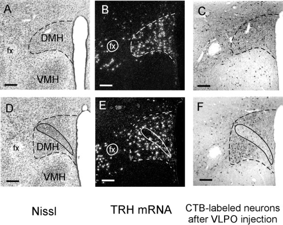
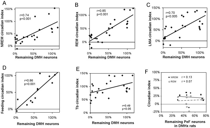
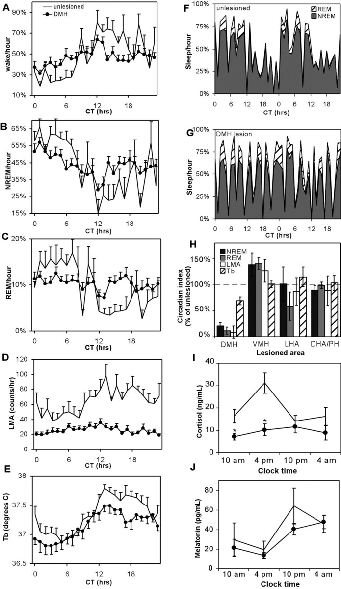
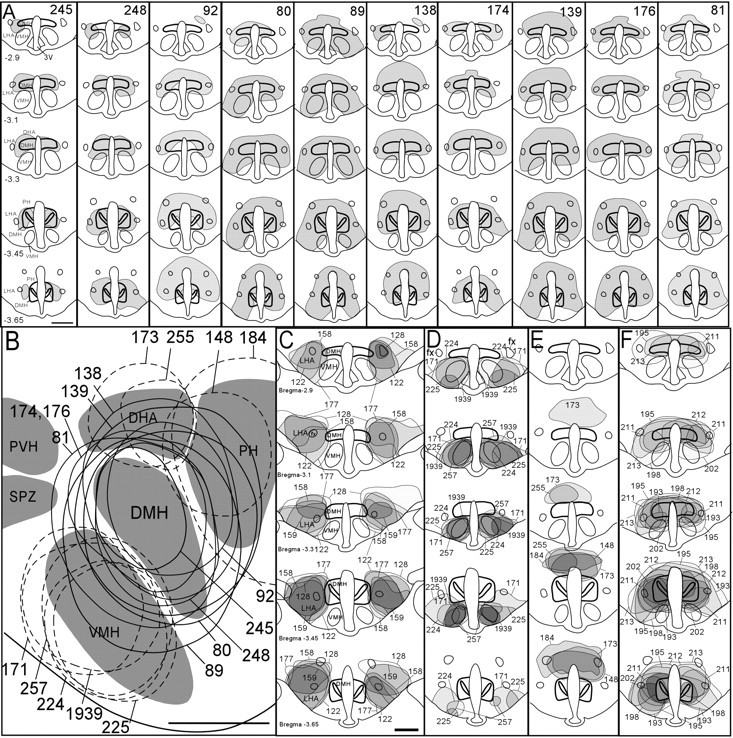
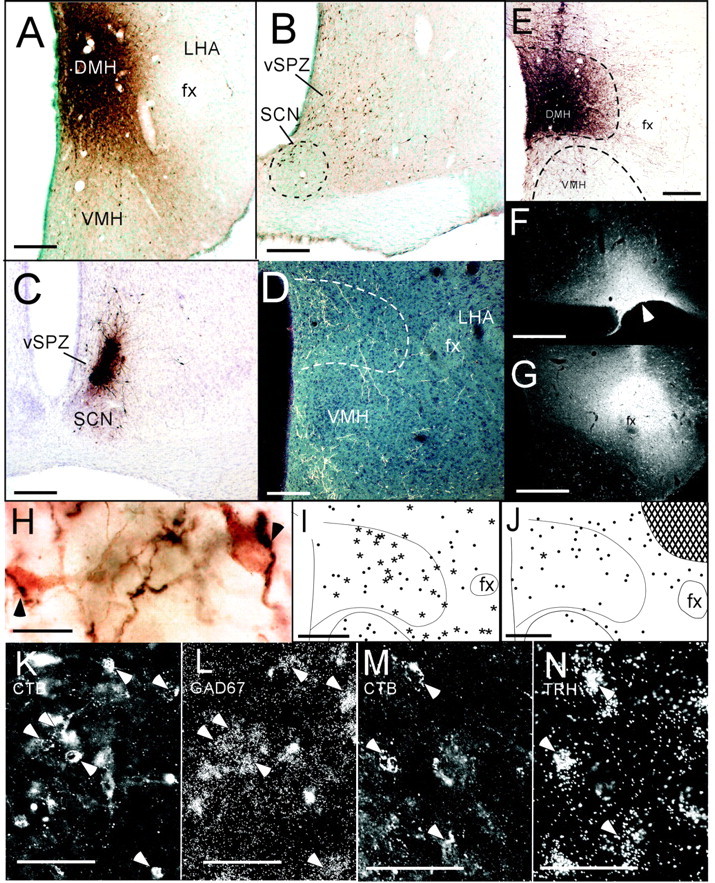
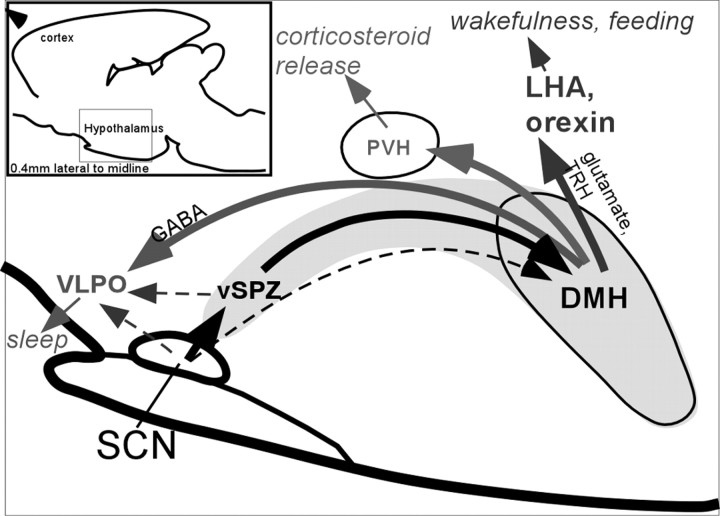
The suprachiasmatic nucleus (SCN) contains the brain's circadian pacemaker, but mechanisms by which it controls circadian rhythms of sleep and related behaviors are poorly understood. Previous anatomic evidence has implicated the dorsomedial hypothalamic nucleus (DMH) in circadian control of sleep, but this hypothesis remains untested. We now show that excitotoxic lesions of the DMH reduce circadian rhythms of wakefulness, feeding, locomotor activity, and serum corticosteroid levels by 78-89% while also reducing their overall daily levels. We also show that the DMH receives both direct and indirect SCN inputs and sends a mainly GABAergic projection to the sleep-promoting ventrolateral preoptic nucleus, and a mainly glutamate-thyrotropin-releasing hormone projection to the wake-promoting lateral hypothalamic area, including orexin (hypocretin) neurons. Through these pathways, the DMH may influence a wide range of behavioral circadian rhythms.


Figures

Figure 1.
Figure 1.
Anatomic markers of DMH borders (dashed lines) in Nissl-stained coronal sections (A) and after in situ hybridization for TRH mRNA (B). Retrograde tracer injections into the VLPO also label many DMH neurons (C). D-F show these same markers at a caudal DMH level where the pars compacta (solid outline) is apparent. Scale bars, 200 μm.
Figure 3.
Figure 3.
After DMH lesions, numbers of surviving DMH neurons correlate significantly with circadian indices of NREM sleep (A), REM sleep (B), LMA (C), and food intake (D), but not Tb (E). The trend lines for sleep, LMA, and feeding, but not Tb, intersect the y-axis near zero, suggesting a greater DMH influence on sleep, LMA, and feeding than Tb rhythms. In DMHx rats (>80% loss of DMH neurons), additional loss of perifornical neurons does not correlate with additional reductions in REM or NREM circadian indices (F). Circadian indices and cell counts are normalized to 100% for sham-operated controls.
Figure 4.
Figure 4.
Hour-by-hour group averages of wakefulness (A), NREM sleep (B), REM sleep (C), and LMA (D) show marked reduction of rhythms in DMHx rats (•), relative to sham-operated controls (no symbol). Results are plotted with respect to circadian time, with the subjective day onset designated CT0. Unlike other rhythms, the Tb circadian rhythm amplitude does not show a marked attenuation (E). Forty-eight consecutive hours of sleep data from a single typical sham-operated control rat show robust NREM and REM circadian rhythms (F), whereas a typical DMH lesioned rat (case 248) shows predominant 3-4 hr ultradian rhythms instead (G). H, Lesions of the VMH, LHA, or DHA/PH show no change, or a modest increase, in circadian indices of sleep, Tb, and LMA, contrasting sharply with DMH-lesioned rats, which have marked reductions of sleep and LMA circadian indices (controls are normalized to 100%). I, DMH area lesions abolish the circadian rhythm of serum cortisol and lower its overall levels but do not affect serum melatonin levels (J). Asterisks in I and J indicate significant differences from sham controls: *p < 0.05; **p < 0.01.
Figure 2.
Figure 2.
A, Coronal views of DMH area lesions ablating >80% of DMH neurons. Drawings are shown in order from least (case 245) to greatest (case 81) cell loss. B, Reconstructions of DMH lesions (solid outlines) in a single sagittal plane 0.4 mm lateral to midline. Also shown are sagittal reconstructions of control lesions ventral, dorsal, or caudal to the DMH (dashed outlines). Rostral is toward the left, and dorsal is toward the top of panel. Lesions lateral to the DMH are not shown in this panel because they lie outside the plane shown. C-E, Coronal drawings of control lesions lateral (C), ventral (D), and dorsal (E) to the DMH. F, Coronal drawings of DMH lesions used for melatonin/corticosteroid measurements. Scale bars, 1 mm. Distance from bregma is indicated in millimeters for coronal sections in A and C.
Figure 5.
Figure 5.
After retrograde tracer injections into the DMH (A), numerous retrogradely labeled cells are seen in the ventral SPZ, with somewhat fewer cells in the dorsomedial SCN (B). Anterograde tracer injections into the ventral SPZ (C) produce labeled terminals mainly in the DMH and VMH (D). After injections of the anterograde tracer biotinylated dextran into the DMH (E), labeled fibers heavily innervate the LHA, particularly near the fornix, where many anterogradely labeled fibers (H, dark reaction product) appose orexin neurons (H, lighter reaction product). After CTB injections into the VLPO (F, arrowhead), many DMH neurons are retrogradely labeled (I), and most of these express GAD67 mRNA (asterisks), although some do not (dots). Higher magnification of DMH region shows retrogradely labeled cells (J, epifluorescence illumination) overlaid with silver grains indicating GAD67 mRNA (K, dark-field illumination). Arrowheads indicate double-labeled cells. After CTB injections into the LHA (G), very few CTB-labeled DMH neurons express GAD67 mRNA (single star), and most lack detectable GAD67 mRNA (dots). Hatched area in J shows CTB injection site. Some DMH neurons retrogradely labeled by LHA injections (M) had overlying silver grains indicating TRH mRNA (N). fx, Fornix. Scale bars: A, D, E, G-I, L, 250 μm; B, C, 125 μm; F, 20 μm; J, K, 150 μm; M, N, 100 μm.
Figure 6.
Figure 6.
Sagittal view (0.4 mm from midline) of major DMH pathways regulating circadian timing of sleep, wakefulness, and hormone secretion. Solid lines indicate relatively intense pathways, whereas dotted lines indicate weaker pathways. Other DMH afferents and efferents not examined in this study are omitted.

References

    1. Abrahamson EE, Leak RK, Moore RY ( 2001) The suprachiasmatic nucleus projects to posterior hypothalamic arousal systems. NeuroReport 12: 435-440. - PubMed
    1. Alfoldi P, Franken P, Tobler I, Borbely AA ( 1991) Short light-dark cycles influence sleep stages and EEG power spectra in the rat. Behav Brain Res 43: 125-131. - PubMed
    1. Amaral DG, Price JL ( 1983) An air pressure system for the injection of tracer substances into the brain. J Neurosci Methods 9: 35-43. - PubMed
    1. Antonio Martinez J, Vargas ML, Fuente T, Del Rio Garcia J, Milanes MV ( 1990) Plasma beta-endorphin and cortisol levels in morphine-tolerant rats and in naloxone-induced withdrawal. Eur J Pharmacol 182: 117-123. - PubMed
    1. Aston-Jones G, Chen S, Zhu Y, Oshinsky ML ( 2001) A neural circuit for circadian regulation of arousal. Nat Neurosci 4: 732-738. - PubMed

Publication types

MeSH terms