

Accommodation of a highly symmetric core within a symmetric protein superfold
- PMID: 14627732
- PMCID: PMC2366980
- DOI: 10.1110/ps.03374903
Accommodation of a highly symmetric core within a symmetric protein superfold
Abstract
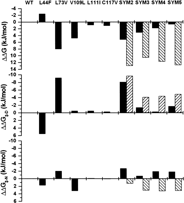
An alternative core packing group, involving a set of five positions, has been introduced into human acidic FGF-1. This alternative group was designed so as to constrain the primary structure within the core region to the same threefold symmetry present in the tertiary structure of the protein fold (the beta-trefoil superfold). The alternative core is essentially indistinguishable from the WT core with regard to structure, stability, and folding kinetics. The results show that the beta-trefoil superfold is compatible with a threefold symmetric constraint on the core region, as might be the case if the superfold arose as a result of gene duplication/fusion events. Furthermore, this new core arrangement can form the basis of a structural "building block" that can greatly simplify the de novo design of beta-trefoil proteins by using symmetric structural complementarity. Remaining asymmetry within the core appears to be related to asymmetry in the tertiary structure associated with receptor and heparin binding functionality of the growth factor.
Figures


Similar articles
-
Redesigning symmetry-related "mini-core" regions of FGF-1 to increase primary structure symmetry: thermodynamic and functional consequences of structural symmetry.Protein Sci. 2005 Sep;14(9):2315-23. doi: 10.1110/ps.051494405. Epub 2005 Aug 4. Protein Sci. 2005. PMID: 16081654 Free PMC article.
-
Symmetric primary and tertiary structure mutations within a symmetric superfold: a solution, not a constraint, to achieve a foldable polypeptide.J Mol Biol. 2004 Nov 26;344(3):769-80. doi: 10.1016/j.jmb.2004.09.060. J Mol Biol. 2004. PMID: 15533444
-
Structure and stability effects of mutations designed to increase the primary sequence symmetry within the core region of a beta-trefoil.Protein Sci. 2001 Dec;10(12):2587-99. doi: 10.1110/ps.ps.34701. Protein Sci. 2001. PMID: 11714927 Free PMC article.
-
Symmetric protein architecture in protein design: top-down symmetric deconstruction.Methods Mol Biol. 2014;1216:161-82. doi: 10.1007/978-1-4939-1486-9_8. Methods Mol Biol. 2014. PMID: 25213415 Review.
-
Emergence of symmetric protein architecture from a simple peptide motif: evolutionary models.Cell Mol Life Sci. 2012 Dec;69(23):3999-4006. doi: 10.1007/s00018-012-1077-3. Epub 2012 Jul 13. Cell Mol Life Sci. 2012. PMID: 22790181 Free PMC article. Review.
Cited by
-
Cooperative hydrophobic core interactions in the β-trefoil architecture.Protein Sci. 2021 May;30(5):956-965. doi: 10.1002/pro.4059. Epub 2021 Mar 16. Protein Sci. 2021. PMID: 33686691 Free PMC article.
-
Redesigning symmetry-related "mini-core" regions of FGF-1 to increase primary structure symmetry: thermodynamic and functional consequences of structural symmetry.Protein Sci. 2005 Sep;14(9):2315-23. doi: 10.1110/ps.051494405. Epub 2005 Aug 4. Protein Sci. 2005. PMID: 16081654 Free PMC article.
-
Experimental support for the foldability-function tradeoff hypothesis: segregation of the folding nucleus and functional regions in fibroblast growth factor-1.Protein Sci. 2012 Dec;21(12):1911-20. doi: 10.1002/pro.2175. Epub 2012 Nov 6. Protein Sci. 2012. PMID: 23047594 Free PMC article.
-
Pharmacokinetic properties of 2nd-generation fibroblast growth factor-1 mutants for therapeutic application.PLoS One. 2012;7(11):e48210. doi: 10.1371/journal.pone.0048210. Epub 2012 Nov 1. PLoS One. 2012. PMID: 23133616 Free PMC article.
-
Experimental support for the evolution of symmetric protein architecture from a simple peptide motif.Proc Natl Acad Sci U S A. 2011 Jan 4;108(1):126-30. doi: 10.1073/pnas.1015032108. Epub 2010 Dec 20. Proc Natl Acad Sci U S A. 2011. PMID: 21173271 Free PMC article.
References
-
- Betz, S.F. and DeGrado, W.F. 1996. Controlling topology and native-like behavior of de novo-designed peptides: Design and characterization of antiparallel four-stranded coiled coils. Biochemistry 35 6955–6962. - PubMed
-
- Betz, S.F., Liebman, P.A., and DeGrado, W.F. 1997. De novo design of native proteins: Characterization of proteins intended to fold into antiparallel, rop-like, four-helix bundles. Biochemistry 36 2450–2458. - PubMed
-
- Blaber, M., DiSalvo, J., and Thomas, K.A. 1996. X-ray crystal structure of human acidic fibroblast growth factor. Biochemistry 35 2086–2094. - PubMed
Publication types
MeSH terms
Substances
Grants and funding
LinkOut - more resources
Full Text Sources
Other Literature Sources
