. 2003 Dec 1;31(23):6689-99.
doi: 10.1093/nar/gkg890.

Sensing DNA damage by PARP-like fingers

Affiliations

Sensing DNA damage by PARP-like fingers

Stefania Petrucco. Nucleic Acids Res. .

Abstract

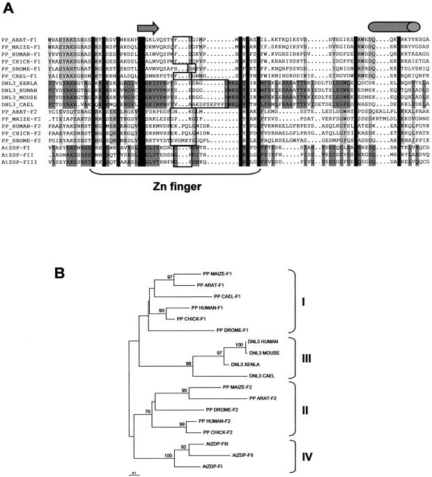
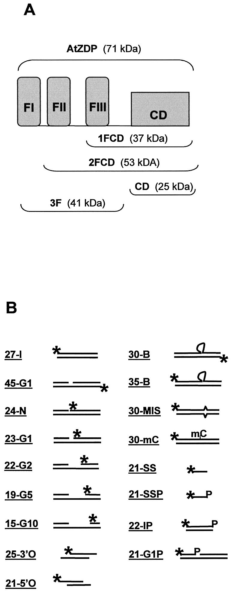
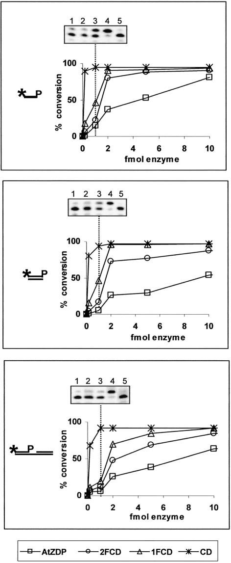
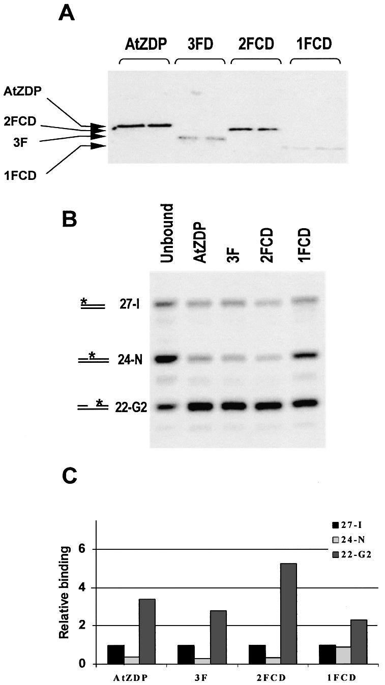
PARP-like zinc fingers are protein modules, initially described as nick-sensors of poly(ADP-ribosyl)-polymerases (PARPs), which are found at the N-terminus of different DNA repair enzymes. I chose to study the role of PARP-like fingers in AtZDP, a 3' DNA phosphoesterase, which is the only known enzyme provided with three such finger domains. Here I show that PARP-like fingers can maintain AtZDP onto damaged DNA sites without interfering with its DNA end repair functions. Damage recognition by AtZDP fingers, in fact, relies on the presence of flexible joints within double-strand DNA and does not entail DNA ends. A single AtZDP finger is already capable of specific recognition. Two fingers strengthen the binding and extend the contacts on the bound DNA. A third finger further enhances the specific binding to damaged DNA sites. Unexpectedly, gaps but not nicks are bound by AtZDP fingers, suggesting that nicks on a naked DNA template do not provide enough flexibility for the recognition. Altogether these results indicate that AtZDP PARP-like fingers, might have a role in positioning the enzyme at sites of enhanced helical flexibility, where single-strand DNA breaks are present or are prone to occur.


Figures

Figure 1
Figure 1
Sequence comparison of PARP-like zinc fingers. (A) Alignment of known PARP-like finger modules. Amino acid residues conserved between all PARP-like finger domains are in black, and amino acids conserved within the clusters identified by the phylogenetic analysis shown in (B) are shaded. Gaps introduced to optimise the alignment are indicated by dots. Sequences within the zinc-coordinating residues are indicated as Zn finger. The arrow and the cylinder indicate respectively, the β-sheet and the α-helical secondary structures, which are predicted to be conserved between all the PARP-like finger domains. The amino acid residues included in a conserved turn, which might indicate the fingertip, are boxed. (B) Phylogenetic relationship among PARP-like finger domains. The tree was generated from the alignment shown in (A) using the neighbour-joining algorithm, and it was rooted with the mid-point distance method. Branches are drawn to scale as indicated by the scale bar, which corresponds to 0.1 changes per site. When >70%, bootstrap percentages are indicated at the nodes. Sequences clusters corresponding to distinct PARP-like finger types are included in brackets.
Figure 2
Figure 2
Structure of protein constructs and of oligo DNAs used in this work. (A) Schematic representation of the AtZDP deletion mutants. The three N-terminal PARP-like fingers (FI, FII, FIII, respectively) and the associated catalytic domain (CD) are shown as closed boxes. The modular composition of the wild type (above) and of the deletion mutants (below) are indicated with corresponding molecular weights enclosed in brackets. (B) Structure of the substrate oligonucleotides. The positions of 32P-labelled 5′-termini (asterisks) and of 3′-phosphate groups (P) are indicated. Heading numbers in oligo names refer to the length in bases of the labelled species. The 45-G1, 24-N, 23-G1, 22-G2, 19-G5, 21G1P are identical oligo- duplexes, only differing for the presence of a nick (24-N) or of a 1-base (45-G1, 23-G1, 21G1P), 2-base (22-G2), 5-base (19-G5), 10-base (15-G10) gap; the 25-3′O and the 21-5′O have overlapping double-strand domains with respectively 3′ or 5′ single-strand extensions on both strands; the 30-B, 35-B, 30-MIS, 30-mC only differ for the presence of a 5-nucleotide bulge (30-B and 35-B), a mismatched (30-MIS) or a methyl-cytosine (30-mC; see Materials and Methods for more details)
Figure 3
Figure 3
DNA 3′-phosphatase activities of AtZDP proteins. The percentages of single-stranded (21-SSP, upper panel), double-stranded (22-IP, middle panel) or of a gapped duplex (21-G1P, lower panel) substrate conversion are plotted against enzyme quantities. All reactions contained 100 fmol of the 5′ labelled-substrate. Unphosphorylated reaction products were separated from phosphorylated DNA substrates on denaturing 8% polyacrylamide gels, quantitated on phosphorimages, and used to calculate the percent of substrate conversion. Data are the average of two independent experiments that differed by <10% of the mean. Symbols are as follows: wild type AtZDP, squares; 2FCD, circles; 1FCD, triangles; CD, asterisks. Phosphorimages of representative phosphatase assays using 1 fmol of AtZDP (lane 1), 2FCD (lane 2), 1FCD (lane 3), CD (lane 4) or 100 fmol of 3F (lane 5) are reported above each graphic. Note that the phosphate-terminated oligonucleotide migrates faster than the hydroxyl-terminated oligonucleotide of the same size.
Figure 4
Figure 4
Interactions between the PARP-like fingers and the DNA 3′-phosphesterase domain modulate AtZDP DNA binding. (A) The DNA binding activity of AtZDP constructs. Phosphorimages of protein–DNA complexes analysed by non-denaturing polyacrylamide gel electrophoresis. 100 fmol of the 32P-labelled 45-G1 gapped duplex (see Fig. 2B) were incubated with the indicated amounts of AtZDP (lanes 2 and 3), 3F (lanes 4 and 5), 2FCD (lanes 6 and 7), 1FCD (lanes 8 and 9) or CD (lanes 10 and 11). The labelled DNA ligand incubated without proteins was run in lane 1. (B) DTT-dependence of AtZDP DNA binding. Phosphorimages of protein–DNA complexes were analysed as in (A) after incubation of 1 pmol of AtZDP (lanes 2 and 3), 3FD (lanes 4 and 5) or 2FCD (lanes 6 and 7) with 100 fmol of the 32P-labelled 45-G1 gapped duplex, either in the absence (lanes 2, 4 and 6) or in the presence (lanes 3, 5 and 7) of DTT. The labelled DNA ligand incubated without protein was run in lane 1.
Figure 5
Figure 5
Oligo-selection analysis in membrane-bound protein–DNA complexes. (A) Phosphorimage of southwestern blotting of fingered AtZDP proteins. Purified proteins were run on an SDS–polyacrylamide gel, transferred to a nitrocellulose membrane and incubated with the 32P-labelled gapped 45-G1 oligo duplex. The migration positions of purified AtZDP proteins loaded on the gel and visualised on the filter after gel transfer are indicated. (B) Phosphorimage of oligo selected by AtZDP proteins. Filter-bound AtZDP proteins were incubated with an equimolar mixture of the 32P-labelled intact 27-I, nicked 24-N and gapped 22-G2 oligo duplexes. After elution from the filter, same amounts of bound radioactivity were analysed by denaturing polyacrylamide gel electrophoresis, as indicated on top of the figure. A sample of the unbound oligo-mixture, recovered after the hybridisation, was run alongside for comparison (Unbound). The migration positions of the labelled species corresponding to the 27-I, the 24-N and the 22-G2 oligo duplexes are indicated on the left. (C) DNA binding preferences by AtZDP constructs. The phosphorimage presented in (B) was used to calculate the relative amount of templates selected by each of the AtZDP constructs. The radioactivity associated with bound oligo 27-I was taken as a reference and given an arbitrary value of 1.
Figure 6
Figure 6
Gap sensing by AtZDP PARP-like fingers. Phosphorimage of labelled oligos, following AtZDP or 3F oligo-selection analyses. DNA templates recovered from filter-bound proteins or from the unbound fraction, as reported in the figure, were identified according to their migration position on denaturing acrylammide gels, and are indicated on the left. (A) Binding to overhanging DNA templates. Overhanging 25-3′O and 21-5′O oligo duplexes, along with the blunt 27-I oligo duplex, were used for the oligo-selection analysis. A quantification of protein-bound oligos relative to bound 27-I duplex is given on the right. (B) AtZDP finger binding to variously gapped DNAs. An equimolar mixture of a 1 (23-G1), 5 (19-G5) and 10 (15-G10) nucleotides-gap oligo duplexes was analysed. Binding ratios are given relative to the amount of bound 23-G1 gapped oligo duplex.
Figure 7
Figure 7
PARP-like fingers sense flexible joints on double-stranded DNA. Phosphorimages of labelled oligos left unbound or recovered from filter-bound 3F, after the incubation with equimolar mixtures of 35-B and 27-I (upper panel), 30-MIS and 27-I (middle panel) or 30-mC and 27-I (lower panel). The migration position of labelled species, and the amounts of 3F-bound oligos relative to the 27-I duplex are indicated on the left and right side, respectively.
Figure 8
Figure 8
AtZDP finger-DNA contact sites. Phosphorimages (A), (B) and (C) and a schematic summary (D) of the DNase I footprint analyses using the 35-B (A), the 30-B (B) or the 45-G1 (C) labelled probe and the indicated AtZDP proteins. A digestion of the protein-free probes (Free) was run alongside for comparison. Nucleotide-numbers are reported relative to the 5′-labelled ends of the corresponding probe. DNA regions protected by AtZDP, 3F and 2FCD are indicated within longer brackets, while shorter brackets delimit the protection by 1FCD. The bulge or gap site is indicated by parentheses on the right side of the phosphorimages. Dashed lines in (D) indicate the footprint extension, which is observed upon a 5-nucleotide extension of the gap.
Figure 9
Figure 9
Schematic view of AtZDP binding to a flexible joint on the DNA. DNA filaments are represented as a ladder, with arrowheads representing the 3′ DNA ends. The bulged or gapped strand is in black. Atoms of the nucleotides contacted by the FIII and the FII fingers are presented using the ball and stick format and are coloured in yellow and pink, respectively. Putative positions of the CD and of the FI finger domains are also suggested.

Similar articles

Cited by

References

    1. Gradwohl G., Menissier de Murcia,J.M., Molinete,M., Simonin,F., Koken,M., Hoeijmakers,J.H. and de Murcia,G. (1990) The second zinc-finger domain of poly(ADP-ribose) polymerase determines specificity for single-stranded breaks in DNA. Proc. Natl Acad. Sci. USA, 87, 2990–2994. - PMC - PubMed
    1. Ikejima M., Noguchi,S., Yamashita,R., Ogura,T., Sugimura,T., Gill,D.M. and Miwa,M. (1990) The zinc fingers of human poly(ADP-ribose) polymerase are differentially required for the recognition of DNA breaks and nicks and the consequent enzyme activation. Other structures recognize intact DNA. J. Biol. Chem., 265, 21907–21913. - PubMed
    1. Caldecott K.W., Aoufouchi,S., Johnson,P. and Shall,S. (1996) XRCC1 polypeptide interacts with DNA polymerase beta and possibly poly (ADP-ribose) polymerase and DNA ligase III is a novel molecular ‘nick-sensor’ in vitro. Nucleic Acids Res., 24, 4387–4394. - PMC - PubMed
    1. Mackey Z.B., Niedergang,C., Murcia,J.M., Leppard,J., Au,K., Chen,J., de Murcia,G. and Tomkinson,A.E. (1999) DNA ligase III is recruited to DNA strand breaks by a zinc finger motif homologous to that of poly(ADP-ribose) polymerase. Identification of two functionally distinct DNA binding regions within DNA ligase III. J. Biol. Chem., 274, 21679–21687. - PubMed
    1. D'Amours D., Desnoyers,S., D’Silva,I. and Poirier,G.G. (1999) Poly(ADP-ribosyl)ation reactions in the regulation of nuclear functions. Biochem. J., 342, 249–268. - PMC - PubMed

Publication types