. 2003 Dec 1;31(23):6806-18.
doi: 10.1093/nar/gkg908.

The common and the distinctive features of the bulged-G motif based on a 1.04 A resolution RNA structure

Affiliations

The common and the distinctive features of the bulged-G motif based on a 1.04 A resolution RNA structure

Carl C Correll et al. Nucleic Acids Res. .

Abstract

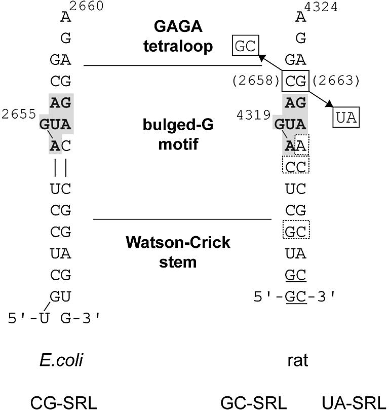
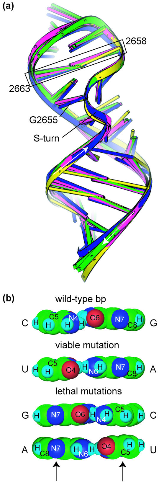
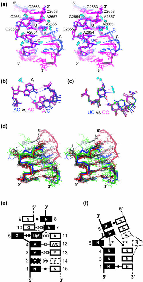
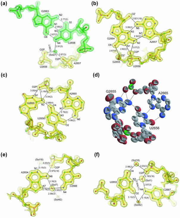
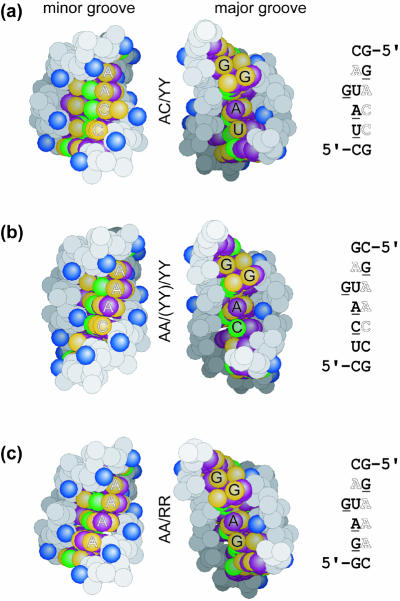
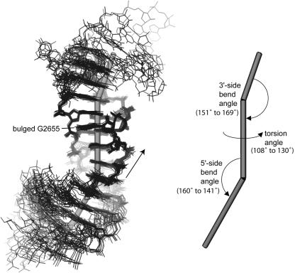
Bulged-G motifs are ubiquitous internal RNA loops that provide specific recognition sites for proteins and RNAs. To establish the common and distinctive features of the motif we determined the structures of three variants and compared them with related structures. The variants are 27-nt mimics of the sarcin/ricin loop (SRL) from Escherichia coli 23S ribosomal RNA that is an essential part of the binding site for elongation factors (EFs). The wild-type SRL has now been determined at 1.04 A resolution, supplementing data obtained before at 1.11 A and allowing the first calculation of coordinate error for an RNA motif. The other two structures, having a viable (C2658U*G2663A) or a lethal mutation (C2658G*G2663C), were determined at 1.75 and 2.25 A resolution, respectively. Comparisons reveal that bulged-G motifs have a common hydration and geometry, with flexible junctions at flanking structural elements. Six conserved nucleotides preserve the fold of the motif; the remaining seven to nine vary in sequence and alter contacts in both grooves. Differences between accessible functional groups of the lethal mutation and those of the viable mutation and wild-type SRL may account for the impaired elongation factor binding to ribosomes with the C2658G*G2663C mutation and may underlie the lethal phenotype.


Figures

Figure 1
Figure 1
Sequences used for crystallization of the E.coli CG-SRL (left), and rat GC-SRL and UA-SRL sequences (right), showing the six conserved nucleotides of the bulged-G motif (gray box). The terminal nucleotides in the rat sequence are designed to form a paired stem (underlined). Mutations of the CG bp in the rat 28S rRNA sequence (E.coli 23S rRNA numbering in parentheses) are boxed. Differences between the bulged-G motif of the E.coli and rat sequences are shown (dashed boxes on the rat sequence).
Figure 2
Figure 2
Structural differences between C2658GG2663C and C2658UG2663A mutations. (a) Superposition of CG-SRL (blue), GC-SRL (green), UA-SRL (magenta) and wild-type rat SRL RNA [yellow; PDB entry 480D (19)]. The mutated bp is boxed, and the bulged G and the S-turn are shown. Arrows indicate the 5′→3′ direction of the phosphodiester backbone. (b) Space-filling models of the Hoogsteen edges of the viable and lethal mutations of the C2658G2663 bp; arrows indicate key functional groups that are different in the lethal and the viable mutations.
Figure 3
Figure 3
Comparisons of bulged-G motifs and variants. (a) The common geometry and hydration of bulged-G motifs among the seven bulged-G motif structures determined at better than 2.0 Å (Materials and Methods) as shown in a stereodiagram of the superposition with AC/YY (blue) and the AA/YY/(YY) variants (purple). (b) Superposition of AC (blue) and AA (purple) bps. (c) Superposition of UC (blue) and CC (purple) bps. Common solvent molecules in (a), (b) and (c) are colored light blue. (d) Stereo diagram of superimposed S2 motifs with the backbone highlighted for four representative variants (Table 3 and Materials and Methods): two for S2I (black and red); S2II (blue) and S2III (green). The two examples of S2I S-turns have different geometries in the strand that opposes the S-turn. The S2II and S2III variants have not been previously identified (14). (e) Stacking diagram for bulged-G motifs with the consensus sequence. The termini form WC bps; N is A, C, G or U. Sometimes positions 3 and 13 are purines, not pyrimidines. Positions are numbered as a reference for torsion angle values in Tables 2 and 3. Standard symbols are used to classify the bps (62); a circled W represents water-mediated interactions. Dashed boxes indicate nucleotides present only in certain variants. (f) Stacking diagram for S2 motifs. Positions are numbered as a reference for torsion angle values given in Table 3. Standard symbols are used to classify the bps (62). Dashed boxes indicate nucleotides present only in certain variants. The base at position 7 stacks in one of two ways (asterisk and double asterisk).
Figure 4
Figure 4
Details of a typical bulged-G motif (S1) determined at 1.04 Å resolution. Final refined model of CG-SRL bps superimposed on a 1.04 Å SIGMAA-weighted 2Fo–Fc density map (47) for (a) the C2658G2663 bp, (b) the A2657G2664 bp, (c) the G2655U2656A2665 base triple, (e) the A2654C2666 bp and (f) the U2653C2667 bp, showing hydrogen bond lengths (Å) and standard errors derived from coordinate errors in parentheses (Å). The distance between O2 of 2653 and N4 of 2667 is 2.965 ± 0.019 Å. This interaction is not drawn as a hydrogen bond because the N-H-O angle is not ideal (58). (d) Each atom of the base triple is represented by a thermal ellipsoid (the more elliptical the shape, the greater the anisotropy) with carbon, oxygen, nitrogen and phosphorous atoms colored gray, red, blue and green, respectively; the alternate conformation for G2655 is included.
Figure 5
Figure 5
Sequence-dependent changes on the surfaces of the bulged-G motif (S1). Space-filling presentations of the minor groove (left) and major groove (center) surfaces of the three classes of the motif, with the corresponding sequences (right): (a) AC/YY (CG-SRL), (b) AA/YY/(YY) [from PDB entry 480D (19)] and (c) AA/RR [from PDB entry 1JJ2 (1,10)]. Base hydrogen bond donor (green) and acceptor (yellow) groups are shown, as are base hydrophobic (purple) and 2′-hydroxyl (blue) groups. Underlined and outlined bases in the sequences are labeled in the major and minor grooves, respectively.
Figure 6
Figure 6
Flexible junctions of the bulged-G motif (S1). Superposition of the 19 bulged-G motif structures determined at 2.4 Å resolution or better (Materials and Methods), showing the corresponding vectors of the motif and its flanking structural elements (left) and the resulting bend and torsion angles (right). The 5′→3′ direction of the bulged strand is shown (arrow).

Similar articles

Cited by

References

    1. Ban N., Nissen,P., Hansen,J., Moore,P.B. and Steitz,T.A. (2000) The complete atomic structure of the large ribosomal subunit at 2.4 Å resolution. Science, 289, 905–920. - PubMed
    1. Wimberly B.T., Brodersen,D.E., Clemons,W.M.,Jr, Morgan-Warren,R.J., Carter,A.P., Vonrhein,C., Hartsch,T. and Ramakrishnan,V. (2000) Structure of the 30S ribosomal subunit. Nature, 407, 327–339. - PubMed
    1. Pioletti M., Schlunzen,F., Harms,J., Zarivach,R., Gluhmann,M., Avila,H., Bashan,A., Bartels,H., Auerbach,T., Jacobi,C. et al. (2001) Crystal structures of complexes of the small ribosomal subunit with tetracycline, edeine and IF3. EMBO J., 20, 1829–1839. - PMC - PubMed
    1. Harms J., Schluenzen,F., Zarivach,R., Bashan,A., Gat,S., Agmon,I., Bartels,H., Franceschi,F. and Yonath,A. (2001) High resolution structure of the large ribosomal subunit from a mesophilic eubacterium. Cell, 107, 679–688. - PubMed
    1. Doherty E.A., Batey,R.T., Masquida,B. and Doudna,J.A. (2001) A universal mode of helix packing in RNA. Nature Struct. Biol., 8, 339–343. - PubMed

Publication types