Comparative Study
. 2003 Dec;73(6):1261-70.
doi: 10.1086/379977. Epub 2003 Nov 18.

Array-based comparative genomic hybridization for the genomewide detection of submicroscopic chromosomal abnormalities

Affiliations
Comparative Study

Array-based comparative genomic hybridization for the genomewide detection of submicroscopic chromosomal abnormalities

Lisenka E L M Vissers et al. Am J Hum Genet. 2003 Dec.

Abstract

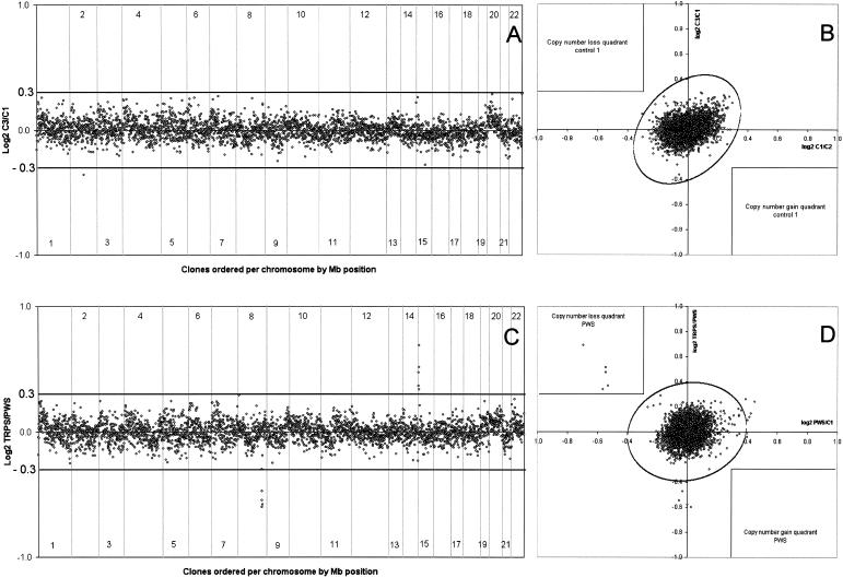
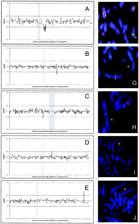
Microdeletions and microduplications, not visible by routine chromosome analysis, are a major cause of human malformation and mental retardation. Novel high-resolution, whole-genome technologies can improve the diagnostic detection rate of these small chromosomal abnormalities. Array-based comparative genomic hybridization allows such a high-resolution screening by hybridizing differentially labeled test and reference DNAs to arrays consisting of thousands of genomic clones. In this study, we tested the diagnostic capacity of this technology using approximately 3,500 flourescent in situ hybridization-verified clones selected to cover the genome with an average of 1 clone per megabase (Mb). The sensitivity and specificity of the technology were tested in normal-versus-normal control experiments and through the screening of patients with known microdeletion syndromes. Subsequently, a series of 20 cytogenetically normal patients with mental retardation and dysmorphisms suggestive of a chromosomal abnormality were analyzed. In this series, three microdeletions and two microduplications were identified and validated. Two of these genomic changes were identified also in one of the parents, indicating that these are large-scale genomic polymorphisms. Deletions and duplications as small as 1 Mb could be reliably detected by our approach. The percentage of false-positive results was reduced to a minimum by use of a dye-swap-replicate analysis, all but eliminating the need for laborious validation experiments and facilitating implementation in a routine diagnostic setting. This high-resolution assay will facilitate the identification of novel genes involved in human mental retardation and/or malformation syndromes and will provide insight into the flexibility and plasticity of the human genome.


Figures

Figure  1
Figure 1
ArrayCGH genomic profiles of validation experiments. Arrays contained 3,343 human autosomal clones (indicated by small circles representing the mean log2-transformed and Lowess-normalized T/R intensity ratios), ordered in A and C from 1pter to 22qter on the basis of the physical mapping positions obtained from the November 2002 freeze of the UCSC genome browser. In panels A and C, chromosome boundaries are indicated by vertical lines. Panel A shows the result of a normal-versus-normal hybridization (control 3 vs. control 1). Nearly all clones fall within the a priori–set thresholds for copy-number gain (log2T/R value 0.3) and copy-number loss (log2 T/R value −0.3) indicated by the horizontal lines. One clone on chromosome 2 shows an intensity ratio outside these thresholds and might represent a false-positive result. Panel B shows the result of the combined analysis of the two hybridizations performed with control 1 (X-axis: control 1 vs. control 2; Y-axis: control 3 vs. control 1). The ellipse represents the border of the reference regions containing 99.999% of the data points; the thresholds for copy-number gain and loss are also integrated into this figure (see the “Patients and Methods” section for details). As can be seen, there is only one clone outside the reference region; however, this clone does not pass the thresholds for copy-number loss in both experiments and can therefore be discarded from further analyses. The clone on chromosome 2 that fell outside the threshold for copy-number loss in panel A is clearly within the normal reference region and can therefore also be discarded for further analyses. Panel C shows the result of the hybridization of DNA from a patient with TRPS against DNA from a patient with PWS. A total of four clones, spanning 2.7 Mb of genomic sequence on 8q23.3-q24.11, showed log2 TRPS-over-PWS intensity ratios below the threshold for copy-number loss, confirming the presence of a deletion of this genomic region in the TRPS patient. In addition, five clones, spanning 2.9 Mb of sequence on 15q11.2, show log2 intensity ratios above the (reverse) threshold for copy-number gain, indicating a deletion of this genomic region in the PWS patient. No clones outside these target genomic regions show potential false-positive results. The combined results of two experiments involving the PWS patient are shown in panel D. The five target clones on 15q11.2 are reproducibly deleted in both experiments and fall outside the bivariate normal distribution reference region (P=.99999) and within the copy-number loss quadrant indicated in the upper left quadrant.
Figure  2
Figure 2
Detailed genomic profiles and FISH validation of copy-number abnormalities identified in five cases with unexplained mental retardation. Panels AE represent individual profiles of the affected chromosomes for each case, with clones ordered for each chromosome from pter to qter on the basis of the physical mapping positions obtained from the November 2002 freeze of the UCSC genome browser. The centromeric region is indicated by a vertical gray dash, the thresholds for copy-number gain (log2 T/R value 0.3) and copy-number loss (log2 T/R value −0.3) are indicated by horizontal lines. Panels FJ represent the FISH validation using (one of) the target clone(s) identified by arrayCGH. Affected chromosomes are indicated by an asterisk (*). Panel A shows the deletion on 7q11 in patient 1, with 14 clones in this region showing an average ratio of −0.5. FISH validation of this case is shown in the adjacent panel F, in which one of the deleted clones on 7q11 is shown in red and a not-deleted control probe is shown in green. Panel B shows the microdeletion on 2q22 in patient 2 with a total of three clones crossing the threshold for copy-number loss, with FISH validation in the adjacent panel G. Deletion of a single clone on 1p21 is shown in panel C for patient 3; this clone was confirmed by FISH to be deleted not only in the patient (panel H) but also in the father of the patient. Copy-number gain detected in a single clone is shown in panels D and E for patients 4 and 5, with FISH confirmation in panels I and J.

References

Electronic-Database Information

    1. BACPAC Resources Center, http://www.chori.org/bacpac/
    1. BACPAC Resources Center's Human BAC Minimal Tiling Set Web site, http://bacpac.chori.org/pHumanMinSet.htm
    1. Online Mendelian Inheritance in Man (OMIM), http://www.ncbi.nlm.nih.gov/Omim/ (for PWS, SMS, and TRPS)

References

    1. Albertson DG, Pinkel D (2003) Genomic microarrays in human genetic disease and cancer. Hum Mol Genet: 12:R145–152 - PubMed
    1. Anderson G, Schroer RJ, Stevenson RE (1996) Mental retardation in South Carolina. II. Causation. In: Saul RA, Phelan MC (eds) Proceedings of the Greenwood Genetic Center. Greenwood Genetic Center, Greenwood, SC, pp 32–44
    1. Ballif BC, Kashork CD, Shaffer LG (2000) The promise and pitfalls of telomere region-specific probes. Am J Hum Genet 67:1356–1359 - PMC - PubMed
    1. Barber JC, Joyce CA, Collinson MN, Nicholson JC, Willatt LR, Dyson HM, Bateman MS, Green AJ, Yates JR, Dennis NR (1998) Duplication of 8p23.1: a cytogenetic anomaly with no established clinical significance. J Med Genet 35:491–496 - PMC - PubMed
    1. Bayes M, Magano LF, Rivera N, Flores R, Perez Jurado LA (2003) Mutational mechanisms of Williams-Beuren syndrome deletions. Am J Hum Genet 73: 131–151 - PMC - PubMed

Publication types