

An over-expression system for characterizing Ppt1 function in Drosophila
- PMID: 14629778
- PMCID: PMC280676
- DOI: 10.1186/1471-2202-4-30
An over-expression system for characterizing Ppt1 function in Drosophila
Abstract
Background: The infantile onset form of Neuronal Ceroid Lipofuscinoses (INCL) is the earliest and most severe form of NCL, with neurological symptoms that reflect massive neurodegeneration in the CNS and retina. INCL is due to recessively inherited mutations at the CLN1 locus. This locus encodes the evolutionarily conserved enzyme palmitoyl-protein thioesterase 1 (PPT1), indicating an essential role for protein palmitoylation in normal neuronal function.
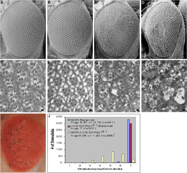
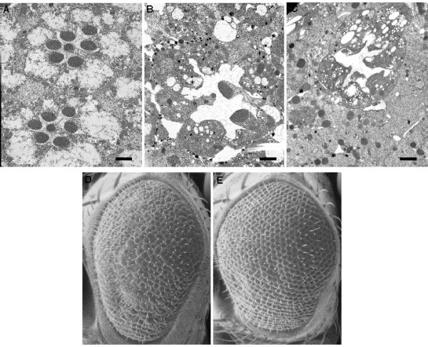
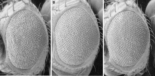
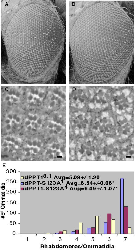
Results: To begin to elucidate the specific role that Ppt1 plays in neuronal cells, we have developed a Ppt1 over-expression system in Drosophila. We report that over-expression of DmPpt1 in the developing Drosophila visual system leads to the loss of cells through apoptotic cell death. This DmPpt1 over-expression phenotype is suppressed by DmPpt1 genomic deficiencies. Moreover, over-expression of DmPpt1S123A, which bears a catalytic site serine 123 to alanine mutation, does not lead to the severe eye phenotype observed with over-expression of wild-type DmPpt1. Thus, cell loss in DmPpt1 flies is directly related to the dosage of wildtype DmPpt1.
Conclusions: Although INCL is due to the loss of PPT1; increased levels of DmPpt1 also lead to neurodegeneration possibly via a detrimental effect on some aspect of PPT1's normal function. This suggests that the precise levels of PPT1 activity are important for neuronal cell survival. The Drosophila DmPpt1 over-expression system provides a resource for genetic experiments that aim to identify the processes by which PPT1 regulates the palmitoylation-state of its essential protein substrates.
Figures
Similar articles
-
Palmitoyl-protein thioesterase 1 deficiency in Drosophila melanogaster causes accumulation of abnormal storage material and reduced life span.Genetics. 2006 Apr;172(4):2379-90. doi: 10.1534/genetics.105.053306. Epub 2006 Feb 1. Genetics. 2006. PMID: 16452138 Free PMC article.
-
Characterization of Drosophila palmitoyl-protein thioesterase 1.Gene. 2003 Jul 17;312:271-9. doi: 10.1016/s0378-1119(03)00623-1. Gene. 2003. PMID: 12909364
-
Glycosylation, transport, and complex formation of palmitoyl protein thioesterase 1 (PPT1)--distinct characteristics in neurons.BMC Cell Biol. 2007 Jun 12;8:22. doi: 10.1186/1471-2121-8-22. BMC Cell Biol. 2007. PMID: 17565660 Free PMC article.
-
Developmental changes in the expression of neuronal ceroid lipofuscinoses-linked proteins.Mol Genet Metab. 2000 Sep-Oct;71(1-2):190-4. doi: 10.1006/mgme.2000.3071. Mol Genet Metab. 2000. PMID: 11001810 Review.
-
Molecular basis of the neuronal ceroid lipofuscinoses: mutations in CLN1, CLN2, CLN3, and CLN5.Hum Mutat. 1999;14(3):199-215. doi: 10.1002/(SICI)1098-1004(1999)14:3<199::AID-HUMU3>3.0.CO;2-A. Hum Mutat. 1999. PMID: 10477428 Review.
Cited by
-
Drosophila melanogaster as a model organism of brain diseases.Int J Mol Sci. 2009 Feb;10(2):407-440. doi: 10.3390/ijms10020407. Epub 2009 Feb 2. Int J Mol Sci. 2009. PMID: 19333415 Free PMC article. Review.
-
Vacuolar protein sorting 35 (Vps35) rescues locomotor deficits and shortened lifespan in Drosophila expressing a Parkinson's disease mutant of Leucine-Rich Repeat Kinase 2 (LRRK2).Mol Neurodegener. 2014 Jun 11;9:23. doi: 10.1186/1750-1326-9-23. Mol Neurodegener. 2014. PMID: 24915984 Free PMC article.
-
The Batten disease gene CLN3 is required for the response to oxidative stress.Hum Mol Genet. 2011 May 15;20(10):2037-47. doi: 10.1093/hmg/ddr088. Epub 2011 Mar 3. Hum Mol Genet. 2011. PMID: 21372148 Free PMC article.
-
Palmitoyl-protein thioesterase 1 deficiency in Drosophila melanogaster causes accumulation of abnormal storage material and reduced life span.Genetics. 2006 Apr;172(4):2379-90. doi: 10.1534/genetics.105.053306. Epub 2006 Feb 1. Genetics. 2006. PMID: 16452138 Free PMC article.
-
Interactions between the juvenile Batten disease gene, CLN3, and the Notch and JNK signalling pathways.Hum Mol Genet. 2009 Feb 15;18(4):667-78. doi: 10.1093/hmg/ddn396. Epub 2008 Nov 21. Hum Mol Genet. 2009. PMID: 19028667 Free PMC article.
References
-
- Rider JA, Rider DL. Batten's Disease: Past, Present, Future. Am J Med Genet Suppl. 1998;5:21–26. - PubMed
-
- Wisniewski KE, Kida E, Golabek AA, Kaczmarski W, Connell F, Zhong N. Neuronal Ceroid Lipofuscinoses: Classification and Diagnosis. Adv Genetics. 2001;45:1–33. - PubMed
-
- Camp LA, Hofmann SL. Purification and Properties of a Palmitoyl-Protein Thioesterase That Cleaves Palmitate From H-Ras. J Biol Chem. 1993;268:22566–22574. - PubMed
Publication types
MeSH terms
Substances
Grants and funding
LinkOut - more resources
Full Text Sources
Molecular Biology Databases
Miscellaneous
