. 2004 Mar 15;103(6):2332-6.
doi: 10.1182/blood-2003-09-3064. Epub 2003 Nov 20.

Characterization of clonogenic multiple myeloma cells

Affiliations

Characterization of clonogenic multiple myeloma cells

William Matsui et al. Blood. .

Abstract

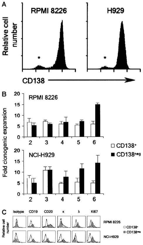
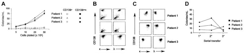
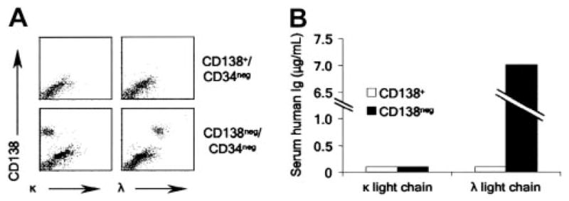
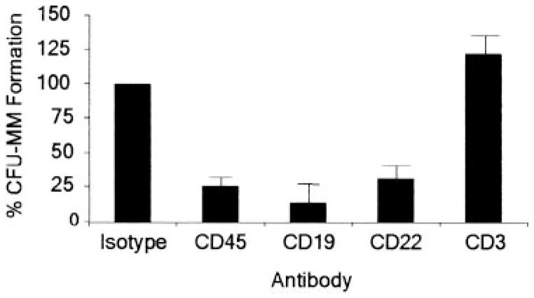
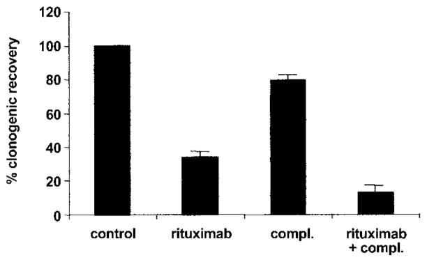
The identity of the cells responsible for the initiation and maintenance of multiple myeloma (MM) remains unclear largely because of the difficulty growing MM cells in vitro and in vivo. MM cell lines and clinical specimens are characterized by malignant plasma cells that express the cell surface antigen syndecan-1 (CD138); however, CD138 expression is limited to terminally differentiated plasma cells during B-cell development. Moreover, circulating B cells that are clonally related to MM plasma cells have been reported in some patients with MM. We found that human MM cell lines contained small (< 5%) subpopulations that lacked CD138 expression and had greater clonogenic potential in vitro than corresponding CD138+ plasma cells. CD138- cells from clinical MM samples were similarly clonogenic both in vitro and in nonobese diabetic/severe combined immunodeficiency (NOD/SCID) mice, whereas CD138+ cells were not. Furthermore, CD138- cells from both cell lines and clinical samples phenotypically resembled postgerminal center B cells, and their clonogenic growth was inhibited by the anti-CD20 monoclonal antibody rituximab. These data suggest that MM "stem cells" are CD138- B cells with the ability to replicate and subsequently differentiate into malignant CD138+ plasma cells.


Figures

Figure 1
Figure 1. Clonogenicity of MM cell lines by CD138 expression
(A) Flow cytometric evaluation of CD138 expression by RPMI 8226 (λ light chain restricted) and NCI-H929 cells (κ light chain restricted). * denotes CD138 population. (B) Clonogenic expansion of CD138+ and CD138 RPMI 8226 and NCI-H929 cells during serial replating. Results are the mean ± SEM of 3 separate experiments for each cell line and represent the fold clonogenic expansion expressed as a ratio of number of colonies scored compared to the previous plating. P < .02 following the fifth and sixth transfer of both cell lines by Student t test. X-axis represents round of serial replating. (C) Flow cytometric evaluation comparing additional antigens expressed by CD138+ and CD138 RPMI 8226 and NCI-H929 cells.
Figure 2
Figure 2. Clonogenic growth of MM cells from clinical specimens
(A) MM colony formation of CD138+ and CD138 cells from 3 clinical samples plated at various densities. (B) Flow cytometric evaluation of colonies derived from CD138/CD34 cells. The top 2 panels show the initial clinical marrow specimen; the middle panels, the CD138-depleted cells prior to plating; and the bottom panels, the pooled colonies after 21 days from a representative MM patient. (C) Flow cytometric evaluation of pooled colonies from 3 additional patients. (D) Colony formation of CD138 clinical MM samples during serial replating.
Figure 3
Figure 3. Engraftment of MM CD138 cells in NOD/SCID mice
(A) Flow cytometric evaluation of murine bone marrow from animals injected with CD138/CD34 or CD138+/CD34 cells from a single patient. (B) Serum human immunoglobulin levels measured by ELISA in mice injected with CD138/CD34 or CD138+/CD34 cells from a single patient.
Figure 4
Figure 4. MM colony formation of CD138/CD34 cells following antibody-mediated cell depletion
Results are presented as a percentage of the untreated control group that was depleted of CD34+ and CD138+ cells. P < .01 for CD45+, CD19+, and CD22+ cell depletions calculated by Student t test comparing each group with the untreated control (n = 12). CFU indicates colony-forming units.
Figure 5
Figure 5. Effect of rituximab on clonogenic MM progenitors in vitro
MM colony formation following treatment of bone marrow mononuclear cells depleted of CD34+ and CD138+ cells with rituximab or 10% human serum (compl.). Results are presented as a percentage of the untreated control group. P < .01 for the rituximab and rituximab plus complement groups compared to the untreated (control) group by Student t test (n = 3).

References

    1. Drewinko B, Alexanian R, Boyer H, Barlogie B, Rubinow SI. The growth fraction of human myeloma cells. Blood. 1981;57:333–338. - PubMed
    1. Hamburger A, Salmon SE. Primary bioassay of human myeloma stem cells. J Clin Invest. 1977;60:846–854. - PMC - PubMed
    1. Pilarski LM, Jensen GS. Monoclonal circulating B cells in multiple myeloma: a continuously differentiating, possibly invasive, population as defined by expression of CD45 isoforms and adhesion molecules. Hematol Oncol Clin North Am. 1992;6:297–322. - PubMed
    1. Billadeau D, Ahmann G, Greipp P, Van Ness B. The bone marrow of multiple myeloma patients contains B cell populations at different stages of differentiation that are clonally related to the malignant plasma cell. J Exp Med. 1993;178:1023–1031. - PMC - PubMed
    1. Bakkus MH, Van RI, Van Camp B, Thielemans K. Evidence that the clonogenic cell in multiple myeloma originates from a pre-switched but somatically mutated B cell. Br J Haematol. 1994;87:68–74. - PubMed

Publication types

MeSH terms