. 2003 Nov 15;17(22):2798-811.
doi: 10.1101/gad.1136103.

Lipoprotein receptors and a disabled family cytoplasmic adaptor protein regulate EGL-17/FGF export in C. elegans

Affiliations

Lipoprotein receptors and a disabled family cytoplasmic adaptor protein regulate EGL-17/FGF export in C. elegans

Darren M Kamikura et al. Genes Dev. .

Abstract

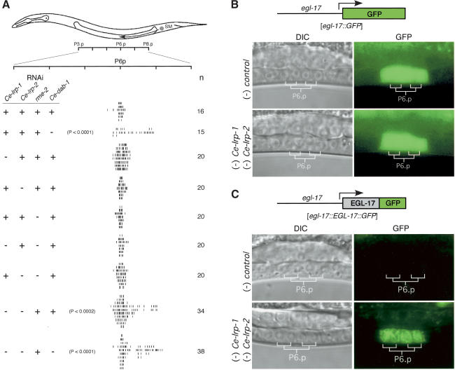
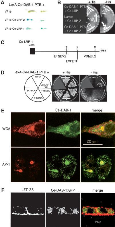
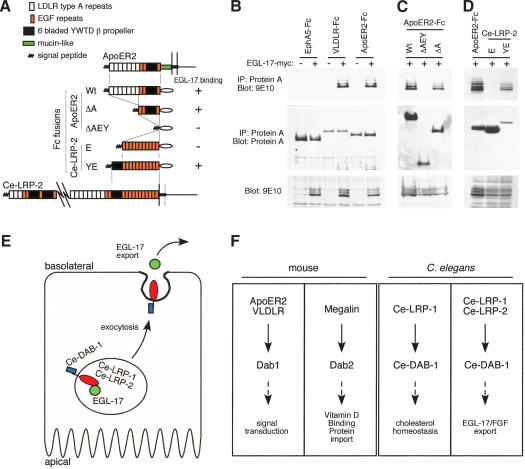
Growth factors and morphogens need to be secreted to act on distant cells during development and in response to injury. Here, we report evidence that efficient export of a fibroblast growth factor (FGF), EGL-17, from the Caenorhabditis elegans developing vulva requires the lipoprotein receptor-related proteins Ce-LRP-1 and Ce-LRP-2 and a cytoplasmic adaptor protein, Ce-DAB-1 (Disabled). Lipoprotein receptors are transmembrane proteins best known for their roles in endocytosis. Ce-LRP-1 and Ce-LRP-2 possess a conserved intraluminal domain that can bind to EGL-17, as well as a cytosolic FXNPXY motif that can bind to Ce-DAB-1. Ce-DAB-1 contains signals that confer subcellular localization to Golgi-proximal vesicles. These results suggest a model in which Ce-DAB-1 coordinates selection of receptors and cargo, including EGL-17, for transport through the secretory pathway.


Figures

Figure 1.
Figure 1.
Characterization of Ce-DAB-1 function and expression. (A) Structural schematic of Ce-DAB-1 protein compared with mouse Dab1 and Dab2 and Drosophila Dab. Blue circles represent NPF sequences and potential binding sites for EH domain proteins. Red triangles represent DPF sequences and potential binding sites for clathrin adaptors. An “X” labels the approximate boundary between 5′ and 3′ portions ofthe cDNA used to generate RNAi. Percentage of identity between the PTB domains relative to Ce-DAB-1 (shaded) is indicated. (BD) RNAi phenotypes. Ce-dab-1 (RNAi) animals exhibit deficiencies in egg laying as adults (B). Black arrowhead indicates an embryo at threefold stage (∼525 cells). Ce-dab-1 (RNAi) animals are also deficient in cuticle molting (C) and resemble lrp-1 RNAi animals (D), exhibiting blisters, “wasp-waist,” and girdle ofunshed cuticle (white arrowhead). (E) Ce-dab-1 (RNAi) disrupts SM migration. The circle labeled “SM” marks the approximate position at which the SMs are generated. The nuclei ofthe Pn.p cells, a set ofhypodermal cells along the ventral side ofthe animal, are used as an anatomical ruler for the measurement of the final positions of the SMs. A vertical hash mark indicates the final position of an individual SM, with an asterisk (*) indicating an individual dorsally localized SM. RNAi used (control or Cedab-1) is indicated in the left column, and the number ofworms scored is indicated in the right column. Statistical significance of altered SM distribution in Ce-dab-1 versus control (RNAi) animals is indicated.
Figure 2.
Figure 2.
Ce-dab-1 is expressed in the developing vulva. (A) Vulvae of qaEx4003[Ce-dab-1 :: Ce-DAB-1 :: GFP; unc-119(+)]; unc-119 (e2498) animals were observed under DIC and fluorescence and photographed as development progressed, increasing in time from top left to bottom right. Before the onset of vulval development (L2; top left panels), expression is seen in the cells of the ventral hypodermis, including the descendants of P5.p, P6.p, and P7.p. As vulval development progresses, expression continues and becomes restricted to the descendants of P5.p and P7.p by the time that vulval invagination is complete (L4; bottom right). Note that the P6.pxxx (great grand-daughters of P6.p) move out of the plane of focus as they adopt their terminal fate. The anchor cell is indicated by an inverted black triangle. Fluorescence above the anchor cell is gut fluorescence. Expression is identical in the independent integrated strain qaIs4000[Ce-dab-1 :: GFP; unc-119(+)]; unc-119(e2498). (B) Ce-dab-1 and egl-17 expression partially overlaps. Schematic illustration of the expression patterns of Ce-dab-1 (green) and egl-17 (red, as described; Burdine et al. 1998) during vulval development.
Figure 3.
Figure 3.
Ce-dab-1 RNAi induces Egl-17—GFP accumulation in the VPCs. (A) Ce-DAB-1 does not regulate egl-17 expression. The onset and level of GFP expression at various stages of vulval development was analyzed in ayIs4[egl-17 :: GFP; dpy-20(+)]; dpy-20(e1282ts) animals (Burdine et al. 1998). Neither control nor Ce-dab-1 RNAi had an effect on the onset of GFP expression driven by the egl-17 promoter. Epifluorescence images were exposed equally. (B) An EGL-17—GFP fusion [egl-17 :: EGL-17 :: GFP] rescues the bag-of-worms phenotype of egl-17(n1377) animals. Progeny of animals expressing the injected pRF4[rol-6(su1006)] coinjection marker were observed for the bag-of-worms phenotype. Fifteen to 30 animals were scored from each set of injections. Empty GFP vector was used as a control. (C) Ce-DAB-1 regulates intracellular distribution of EGL-17 :: GFP. qaEx4002[egl-17 :: EGL-17 :: GFP; unc-119(+)]; unc-119(e2498) transgenic animals were tested for changes in EGL-17 :: GFP distribution after treatment with control or Ce-dab-1 RNAi. Equal exposure times (long or short) were used to record GFP fluorescence. Ce-dab-1 (RNAi) resulted in accumulation of EGL-17—GFP in ∼70% of animals, compared with <5% in controls. A total of >14 independent experiments with >10 animals were performed. Similar results were obtained with the integrated strain qaIs4004[Ce-dab-1 :: Ce-DAB-1 :: GFP; unc-119(+)]; unc-119(e2498). (D) LET-23 and AJM-1 localization is unaltered, whereas EGL-17 :: GFP accumulates in Ce-dab-1 (RNAi) animals. qaEx4002[egl-17 :: EGL-17 :: GFP; unc-119(+)]; unc-119(e2498) animals treated with control or Ce-dab-1 RNAi were analyzed by indirect immunofluorescence for LET-23 and AJM-1 (center panels) or LET-23 and EGL-17 :: GFP (anti-GFP; left and right panels). Left panels show EGL-17 :: GFP in a single 0.2 μm confocal plane of P6.p, whereas right panels show a single confocal plane of P6.p after two divisions. Center panels show a single 0.2-μm confocal plane of P6.p after one division. Bar, 20 μm.
Figure 4.
Figure 4.
Lipoprotein receptors Ce-LRP-1 and Ce-LRP-2, but not RME-2, regulate SM migration. (A) Ce-LRP-1 and Ce-LRP-2 regulate SM migration. The positions ofindividual SMs were measured as in Figure 1. dsRNA was added as indicated: (-) RNAi present; (+) no RNAi. Numbers in the right column indicate the number ofanimals scored. (B) The lipoprotein receptors Ce-LRP-1 and Ce-LRP-2 do not regulate EGL-17 expression. egl-17::GFP animals were analyzed after treatment with control or Ce-lrp-1; Ce-lrp-2 RNAi. (C) Ce-LRP-1 and Ce-LRP-2 regulate EGL-17 export from the VPCs. qaEx4002[egl-17::EGL-17::GFP; unc-119(+)]; unc-119(e2498) transgenic animals were tested for changes in subcellular distribution of the EGL-17::GFP fusion after treatment with control or Ce-lrp-1; Ce-lrp-2 RNAi.
Figure 5.
Figure 5.
Ce-DAB-1 associates with Ce-LRP-1 and Ce-LRP-2 and with Golgi components. (A,B) The Ce-DAB-1 PTB domain (residues 1–252) shows interaction with the intracellular domains ofboth Ce-LRP-1 (residues 4595–4753) and Ce-LRP-2 (C-terminal 81 residues) by yeast two-hybrid assay. β-Galactosidase activity (replicates; A) and growth in the absence ofhistidine (B). (C) Potential binding sites in Ce-LRP-1 (underlined amino acids indicate those that match the consensus binding site for the PTB domains of mammalian Dab1 and Dab2). (D) The Ce-DAB-1 PTB domain interacts with an FXNPXY motiffrom residues 4653–4658. Substitution of Tyr 4658 with alanine reduces interaction between the Ce-DAB-1 PTB domain and the intracellular domain of Ce-LRP-1. (E) Subcellular localization of Ce-DAB-1 expressed in tissue culture cells. NIH3T3 (WGA) and HeLa cells (γ-adaptin) were transfected to express Ce-DAB-1 and processed for indirect immunofluorescence with antibodies to γ-adaptin subunit AP-1, Ce-DAB-1, and directly labeled WGA. Note that there are two HeLa cells in the image shown. (F) Ce-DAB-1::GFP is present in punctate structures within P6.p daughters. Indirect immunofluorescence of qaEx4003 [Ce-dab-1::Ce-DAB-1::GFP; unc-119(+)]; unc-119(e2498) animals with antibodies to LET-23 (left) and Ce-DAB-1::GFP (anti-GFP; center) and merged images (right) are shown. Images are single 0.2-μm confocal planes through P6.p after one division.
Figure 6.
Figure 6.
EGL-17 binds to the extracellular domains oflipoprotein receptors. (AD) 293T cells were transfected with constructs encoding various extracellular domains fused to Fc in the presence or absence of EGL-17-myc. Lysates were precipitated with Protein A Sepharose, resolved by 9% SDS-PAGE, and analyzed by Western blotting. (A) Summary ofconstructs and results. (B) VLDLR and ApoER2, but not EphA5, coprecipitate EGL-17-myc (top). Expression levels of Fc fusion proteins (middle) and EGL-17-myc (bottom) are shown. (C) The association of EGL-17 with ApoER2 is independent ofthe LDLR type A repeats. Expression levels ofthe ApoER2 ΔAEY and ApoER2 ΔA forms (middle) and EGL-17-myc (bottom) are shown. (D) EGL-17 binds to Ce-LRP-2 YWTD region. Despite weaker expression, Ce-LRP-2 YE coprecipitates EGL-17-myc, but Ce-LRP-2 E does not. (E) Model. Ce-DAB-1 regulates the export of EGL-17 mediated through Ce-LRP-1 and Ce-LRP-2, possibly by direct interactions between Ce-DAB-1 in the cytosol, Ce-LRP-1 and Ce-LRP-2 in the membranes ofsecretory vesicles, and EGL-17 in the lumen of the vesicles. See text for discussion. (F) Functions of lipoprotein receptors and Dab family members across species.

Similar articles

Cited by

References

    1. Amanai K. and Jiang, J. 2001. Distinct roles of Central missing and Dispatched in sending the Hedgehog signal. Development 128: 5119-5127. - PubMed
    1. Borland C.Z., Schutzman, J.L., and Stern, M.J. 2001. Fibroblast growth factor signaling in Caenorhabditis elegans. BioEssays 23: 1120-1130. - PubMed
    1. Branda C.S. and Stern, M.J. 2000. Mechanisms controlling sex myoblast migration in Caenorhabditis elegans hermaphrodites. Dev. Biol. 226: 137-151. - PubMed
    1. Burdine R.D., Chen, E.B., Kwok, S.F., and Stern, M.J. 1997. egl-17 encodes an invertebrate fibroblast growth factor family member required specifically for sex myoblast migration in Caenorhabditis elegans. Proc. Natl. Acad. Sci. 94: 2433-2437. - PMC - PubMed
    1. Burdine R.D., Branda, C.S., and Stern, M.J. 1998. EGL-17(FGF) expression coordinates the attraction ofthe migrating sex myoblasts with vulval induction in C. elegans. Development 125: 1083-1093. - PubMed

Publication types

MeSH terms

Substances

LinkOut - more resources