. 2003 Dec;15(12):2911-28.
doi: 10.1105/tpc.017541. Epub 2003 Nov 20.

Poplar metal tolerance protein 1 confers zinc tolerance and is an oligomeric vacuolar zinc transporter with an essential leucine zipper motif

Affiliations

Poplar metal tolerance protein 1 confers zinc tolerance and is an oligomeric vacuolar zinc transporter with an essential leucine zipper motif

Damien Blaudez et al. Plant Cell. 2003 Dec.

Abstract

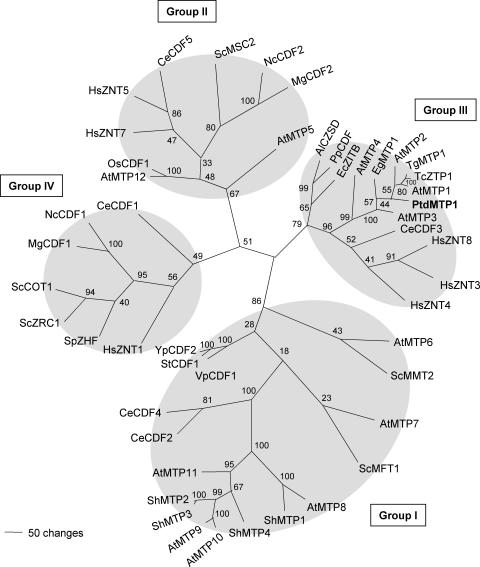
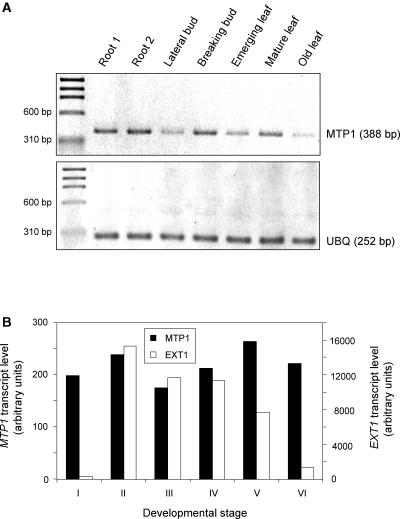
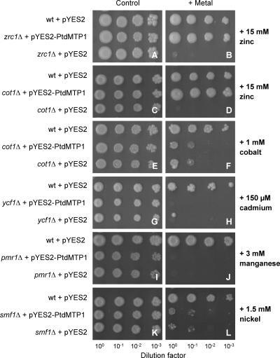
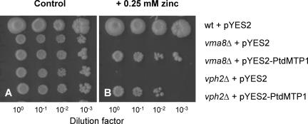
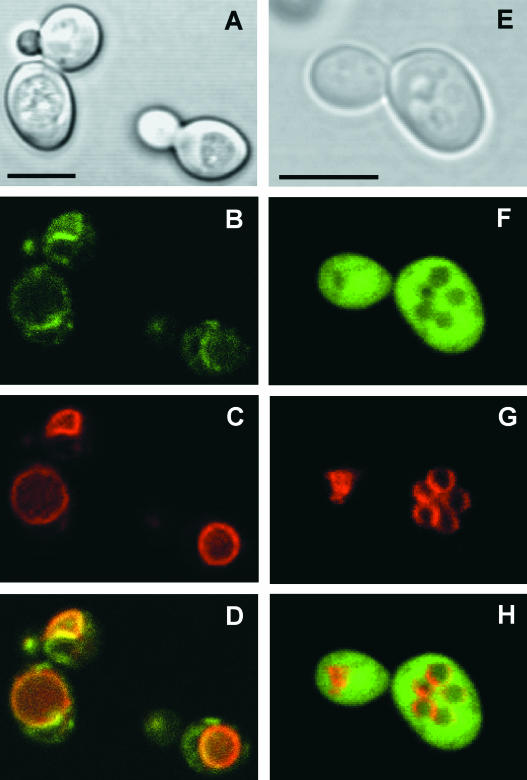
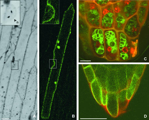
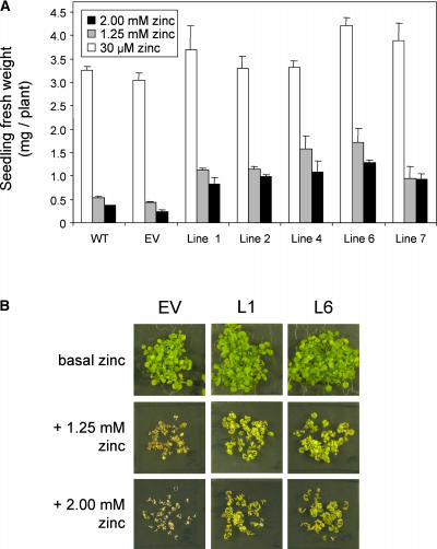
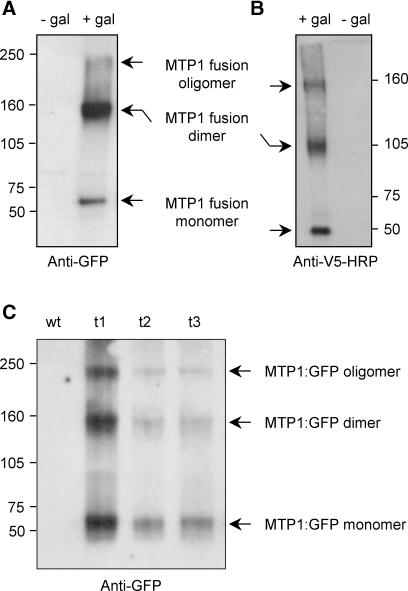
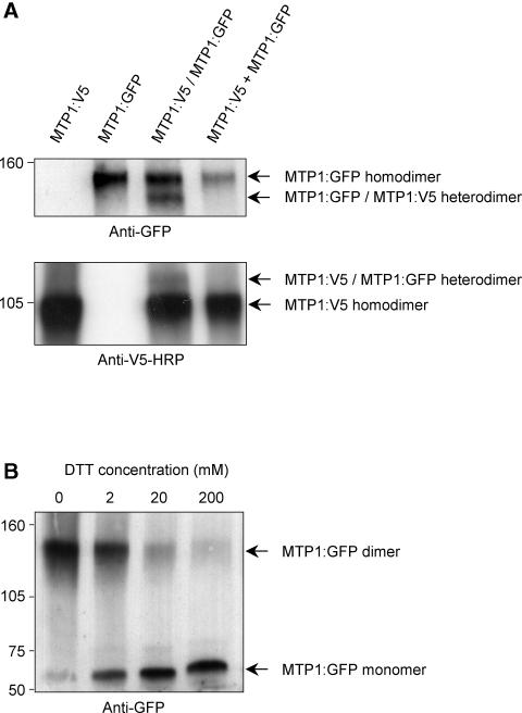
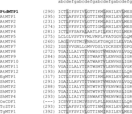
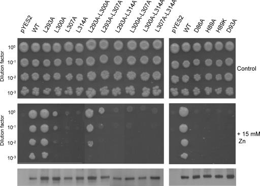
Cation diffusion facilitator (CDF) proteins are a recently discovered family of cation efflux transporters that might play an essential role in metal homeostasis and tolerance. Here, we describe the identification, characterization, and localization of PtdMTP1, a member of the CDF family from the hybrid poplar Populus trichocarpa x Populus deltoides. PtdMTP1 is expressed constitutively and ubiquitously, although at low levels. Heterologous expression in yeast showed that PtdMTP1 was able to complement the hypersensitivity of mutant strains to Zn but not to other metals, including Cd, Co, Mn, and Ni. PtdMTP1 fused to green fluorescent protein localized to the vacuolar membrane both in yeast and in plant cells, consistent with a function of PtdMTP1 in zinc sequestration. Overexpression of PtdMTP1 in Arabidopsis confers Zn tolerance. We show that PtdMTP1, when expressed in yeast and Arabidopsis, forms homooligomers, a novel feature of CDF members. Oligomer formation is disrupted by reducing agents, indicating possible disulfide bridge formation. PtdMTP1 also contains a conserved Leu zipper motif. Although not necessary for oligomer formation, Leu residues within this motif are required for PtdMTP1 functional activity.


Figures

Figure 1.
Figure 1.
An Unrooted, Parsimony-Based Tree of the CDF Gene Family. The tree was generated using PAUP 4.0b10 (D. Swofford, Smithsonian Institution, Washington, DC) after sequence alignment with CLUSTAL X (Thompson et al., 1997). Bootstrap values are indicated (1000 replicates, full heuristic search option). Accession numbers are given at the end of Methods. Arabidopsis gene names follow the new nomenclature used by Delhaize et al. (2003).
Figure 2.
Figure 2.
Expression Levels of PtdMTP1 in Poplar. (A) Transcript levels in different tissues of mature poplar were estimated by reverse transcription–PCR as described in Methods. The expression level of PtdMTP1 was compared with that of UBIQUITIN (UBQ). Experiments were repeated three times, and representative gels are shown. (B) Transcript levels during the development of adventitious poplar roots were estimated from cDNA arrays as described in Methods. Developmental stages are as follows: I, bark tissues of dormant cuttings; II, root primordium; III, root callus; IV, emerging roots; V, primary roots; and VI, lateral root tips. The expression level of PtdMTP1 was compared with that of EXTENSIN (EXT1), a highly expressed and strongly regulated gene. Experiments were performed in triplicate, and representative data are shown.
Figure 3.
Figure 3.
Complementation of Yeast Mutants on Selective Media. S. cerevisiae mutant strains were transformed with the empty vector pYES2 or with pYES2-PtdMTP1. Wild-type (wt) cells (strain BY4741) also were transformed with pYES2 as a control. Yeast cultures were adjusted to OD = 1.0, and 2 μL of serial dilutions (from left to right in each panel) were spotted on SD medium without extra metal ([A], [C], [E], [G], [I], and [K]) or supplemented with 15 mM Zn ([B] and [D]), 1 mM Co (F), 150 μM Cd (H), 3 mM Mn (J), or 1.5 mM Ni (L). Plates were incubated for 6 days at 30°C.
Figure 4.
Figure 4.
Partial complementation of Yeast Vacuolar Acidification Mutants. For yeast transformation and growth conditions, see legend to Figure 3. Yeast growth was assayed on SD medium without extra metal (A) or supplemented with 0.25 mM Zn (B), with serial dilutions from left to right in each panel. wt, wild type.
Figure 5.
Figure 5.
Vacuolar Membrane Localization of the PtdMTP1:GFP Fusion Protein in Yeast, Viewed by Confocal Laser Scanning Microscopy. Cells were visualized after 24 h of induction. Four images from the same cells are shown: bright-field image (A), GFP fluorescence from a PtdMTP1:GFP-expressing strain (B), FM4-64 staining of vacuolar membranes (C), and a pseudocolored merged image with GFP in green and FM4-64 in red (D). (E) to (H) are as for (A) to (D) except for (F) and (H), which show GFP fluorescence from nonfused GFP. Bars = 5 μm.
Figure 6.
Figure 6.
Vacuolar Membrane Localization of PtdMTP1:GFP in Planta. (A) and (B) Transient expression of PtdMTP1:GFP in onion epidermal cells. Bar = 50 μm. (A) Bright-field image along the middle plane of a cell showing the nucleus (n) and invagination of the vacuolar membrane (inset). (B) GFP fluorescence in the same cell concentrated to the vacuolar membrane, which follows the cell contour except in the perinuclear region (inset). (C) and (D) Vacuolar localization of PtdMTP1:GFP in Arabidopsis. The red fluorescence is caused by cell walls and nuclei stained with propidium iodide. Bars = 10 μm. (C) Root tips of transgenic Arabidopsis expressing PtdMTP1:GFP. (D) Control cells expressing AtMGT1:GFP, a transporter located in the plasma membrane.
Figure 7.
Figure 7.
Overexpression of PtdMTP1 Confers Zn Tolerance to Arabidopsis. (A) Plant fresh weight of Arabidopsis lines expressing PtdMTP1 on control half-strength MS medium (containing 30 μM Zn) or supplemented with 1.25 or 2 mM Zn. Plants were harvested after 3 weeks of growth on plates. Lines 1, 2, 4, 6, and 7 are independent homozygous lines expressing PtdMTP1. EV indicates a homozygous line transformed with the empty vector pART27. WT refers to untransformed Arabidopsis wild type. Data are means of 28 to 42 plants, and vertical bars represent standard errors. (B) Zn-tolerance test of two representative Arabidopsis lines expressing PtdMTP1 and a control line (EV) transformed with the empty vector after 3 weeks of growth on control (basal), 1.25 mM, or 2 mM Zn plates.
Figure 8.
Figure 8.
Oligomerization of PtdMTP1 Expressed in Yeast and Arabidopsis. S. cerevisiae expressing either PtdMTP1:GFP (A) or PtdMTP1:V5 (B) were induced or not with 2% (w/v) galactose (gal). (C) shows the expression of PtdMTP1:GFP in Arabidopsis control plants (wt) or three transgenic lines (t1 to t3) overexpressing PtdMTP1:GFP. Proteins were extracted and separated on 5% (w/v) SDS-PAGE gels under nonreducing conditions and analyzed further using immunoblotting methods. The positions of the PtdMTP1:GFP and PtdMTP1:V5 monomers, dimers, and oligomers are indicated. The antibodies used for detection are noted below each gel. Numbers beside the gels indicate molecular masses in kilodaltons.
Figure 9.
Figure 9.
PtdMTP1 Forms Dimers. (A) Coexpression of PtdMTP1:GFP and PtdMTP1:V5 fusion proteins in yeast shows dimer formation. As indicated above lanes 1 to 3 (from left), proteins were extracted from cells expressing only PtdMTP1:V5, only PtdMTP1:GFP, or both (PtdMTP1:GFP/PtdMTP1:V5). The right lane was loaded with proteins from cells expressing only PtdMTP1:GFP mixed with cells expressing only PtdMTP1:V5 (PtdMTP1:GFP + PtdMTP1:V5). (B) Disruption of the PtdMTP1:GFP dimer by DTT. Samples were treated with increasing concentrations of DTT for 1 h before loading. Cells were processed for immunodetection as described in the legend to Figure 8. The positions of homodimers and heterodimers are indicated. The antibodies used for detection are noted below each gel. Numbers beside the gels indicate molecular masses in kilodaltons.
Figure 10.
Figure 10.
Conservation of a LZ Motif in Plant CDFs. Numbers in parentheses correspond to the first residue in each sequence. Gray-shaded residues indicate conserved “d” positions of the Leu/Ile/Val heptad repeats. Sequences were aligned as described in the legend to Figure 1. Accession numbers are given at the end of Methods.
Figure 11.
Figure 11.
Evaluation of the Functionality of PtdMTP1 Mutants by Yeast Complementation Assay. Yeast zrc1Δ mutants were transformed with the empty vector pYES2, with pYES2-PtdMTP1, or with various mutant pYES2-PtdMTP1 vectors. Cultures were adjusted to OD = 1.0, and 2 μL of serial dilutions were spotted on SD medium without extra metal (top panels) or supplemented with 15 mM Zn (middle panels). Plates were incubated for 6 days at 30°C. Protein gel blot analysis of microsomes of transformed mutants with anti-GFP antibodies is shown in the bottom panel. Cells were processed for immunodetection as described in the legend to Figure 8. WT, wild type.

Similar articles

Cited by

References

    1. Altschul, S.F., Gish, W., Miller, W., Myers, E.W., and Lipman, D.J. (1990). Basic local alignment search tool. J. Mol. Biol. 215, 403–410. - PubMed
    1. Bachhawat, A.K., Manolson, M.F., Murdock, D.G., Garman, J.D., and Jones, E.W. (1993). The VPH2 gene encodes a 25 kDa protein required for activity of the yeast vacuolar H+-ATPase. Yeast 9, 175–184. - PubMed
    1. Bloss, T., Clemens, S., and Nies, D.H. (2002). Characterization of the ZAT1p zinc transporter from Arabidopsis thaliana in microbial model organisms and reconstituted proteoliposomes. Planta 214, 783–791. - PubMed
    1. Chardonnens, A.N., Koevoets, P.L., van Zanten, A., Schat, H., and Verkleij, J.A. (1999). Properties of enhanced tonoplast zinc transport in naturally selected zinc-tolerant Silene vulgaris. Plant Physiol. 120, 779–786. - PMC - PubMed
    1. Clemens, S. (2001). Molecular mechanisms of plant metal tolerance and homeostasis. Planta 212, 475–486. - PubMed

Publication types

MeSH terms

Associated data