Review
. 2003 Dec;110(4):411-20.
doi: 10.1111/j.1365-2567.2003.01756.x.

B-cell antigen-receptor signalling in lymphocyte development

Affiliations
Review

B-cell antigen-receptor signalling in lymphocyte development

Leo D Wang et al. Immunology. 2003 Dec.

Abstract

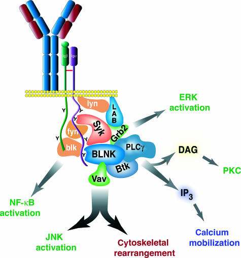
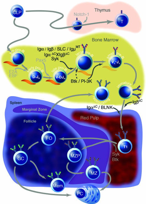
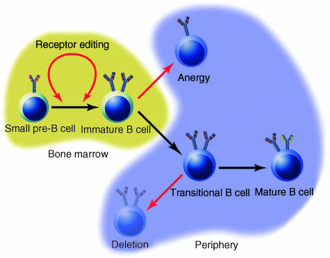
Signalling through the B-cell antigen receptor (BCR) is required throughout B-cell development and peripheral maturation. Targeted disruption of BCR components or downstream effectors indicates that specific signalling mechanisms are preferentially required for central B-cell development, peripheral maturation and repertoire selection. Additionally, the avidity and the context in which antigen is encountered determine both cell fate and differentiation in the periphery. Although the signalling and receptor components required at each stage have been largely elucidated, the molecular mechanisms through which specific signalling are evoked at each stage are still obscure. In particular, it is not known how the pre-BCR initiates the signals required for normal development or how immature B cells regulate the signalling pathways that determine cell fate. In this review, we will summarize the recent studies that have defined the molecules required for B-cell development and maturation as well as the theories on how signals may be regulated at each stage.


Figures

Figure 1
Figure 1
Proximal B-cell receptor-mediated signalling pathways. After binding to antigen, the immunoglobulin (Ig)α and Igβ cytoplasmic tails are phosphorylated on the immunoreceptor tyrosine-based activation motif (ITAM) tyrosines by Src-family tyrosine kinases (SFTKs) and/or Syk. Syk then binds to the Igα ITAM, and the B-cell linker protein (BLNK) binds to tyrosine 204 of Igα. This activates multiple signalling pathways, including: Btk, which activates phospholipase C (PLC)γ2 and leads to calcium flux (blue) and protein kinase C (PKC) activation (green); Grb2, which activates the Ras/Raf/mitogen-activated protein kinase (MEK) extracellular signal-regulated kinase (ERK) pathway (green); and Vav, which activates the Rac/Rho/Cdc42 pathway and results both in cytoskeletal rearrangement (maroon) and c-Jun N-terminal protein kinase (JNK) activation (green). The SFTKs themselves activate nuclear factor-κB (NF-κB) (green).
Figure 2
Figure 2
Overview of B-cell development in postnatal mammalian life. Common lymphoid progenitors (CLPs) can migrate to the thymus where they become T cells in response to signalling through Notch-1, or they can remain in the bone marrow where they enter the B lineage as pre-pro-B cells. Possibly in response to Notch-1 down-regulation by Pax5, transcription factors EBF and E2A are up-regulated to initiate a B-cell-specific programme of gene transcription. DH–JH rearrangements occur at the early pro-B stage, and subsequent V–DJ rearrangements commence at the late pro-B stage. These stages require direct contact with bone marrow stromal cells, depicted in orange. Signalling molecules, whose absence results in a developmental block, are listed in black; dashed arrows indicate incomplete impairment. Appropriate signalling through the pre-B-cell receptor (BCR) (the surrogate light chain is depicted in orange) mediates heavy-chain allelic exclusion and induces several rounds of proliferation. Subsequently, at the small pre-B stage, light-chain rearrangements begin. Successfully rearranged heavy and light chains appear on the cell surface at the immature B-cell stage, after which B cells emigrate to the periphery. Newly immigrated transitional immature B cells (T1) probably undergo further differentiation into T2 and possibly T3 transitional immature B cells, although this is controversial. Eventually, transitional cells enter splenic follicles, where they differentiate further along the marginal zone (MZP, MZ) or follicular (FO) pathways, probably in response to the strength of signals received through the BCR. Follicular cells differentiate either into plasma cells (PCs) or germinal centre B cells (GC) in response to primary antigen challenge, whereupon they undergo somatic hypermutation, affinity maturation and class switch before they differentiate into memory cells. Marginal zone cells, which leave the splenic follicle to reside in the marginal zone, are also capable of differentiating into plasma cells in response to primary antigen exposure.
Figure 3
Figure 3
Mechanisms of negative selection during B-cell development. There are three described mechanisms whereby developing B cells can escape an autoreactive fate; all of these are mediated by signals transduced through the B-cell receptor (BCR) in response to BCR ligation and are indicated by red arrows. Which outcome results is largely a function of BCR signalling intensity, developmental stage and environmental milieu. Receptor editing is postulated to be a result either of retarded progression from the pre-B to immature B-cell stage, or of back-differentiation from the immature to pre-B-cell stage. Anergy is induced in immature bone marrow B cells as a response to receptor ligation, whereas deletion is induced in transitional B cells in response to the same stimulus.

References

    1. Raff MC, Steinberg J, Taylor R. Immunoglobulin determinants on the surface of mouse lymphoid cells. Nature. 1970;225:553. - PubMed
    1. Grupp SA, Campbell K, Mitchell RN, Cambier JC, Abbas AK. Signaling-defective mutants of the B lymphocyte antigen receptor fail to associate with Ig-alpha and Ig-beta/gamma. J Biol Chem. 1993;268:25776. - PubMed
    1. Mitchell RN, Shaw AC, Weaver YK, Leder P, Abbas AK. Cytoplasmic tail deletion converts membrane immunoglobulin to a phosphatidylinositol-linked form lacking signaling and efficient antigen internalization functions. J Biol Chem. 1991;266:8856. - PubMed
    1. Blum JH, Stevens TL, Defranco AL. Role of the mu immunoglobulin heavy chain transmembrane and cytoplasmic domains in B-cell antigen receptor expression and signal transduction. J Biol Chem. 1993;268:27236. - PubMed
    1. Martin SW, Goodnow CC. Burst-enhancing role of the IgG membrane tail as a molecular determinant of memory. Nat Immunol. 2002;3:182. - PubMed

MeSH terms

LinkOut - more resources