. 2003 Nov;110(3):296-303.
doi: 10.1046/j.1365-2567.2003.01745.x.

In vivo expression of interleukin-8, and regulated on activation, normal, T-cell expressed, and secreted, by human germinal centre B lymphocytes

Affiliations

In vivo expression of interleukin-8, and regulated on activation, normal, T-cell expressed, and secreted, by human germinal centre B lymphocytes

Jennifer C Sims-Mourtada et al. Immunology. 2003 Nov.

Abstract

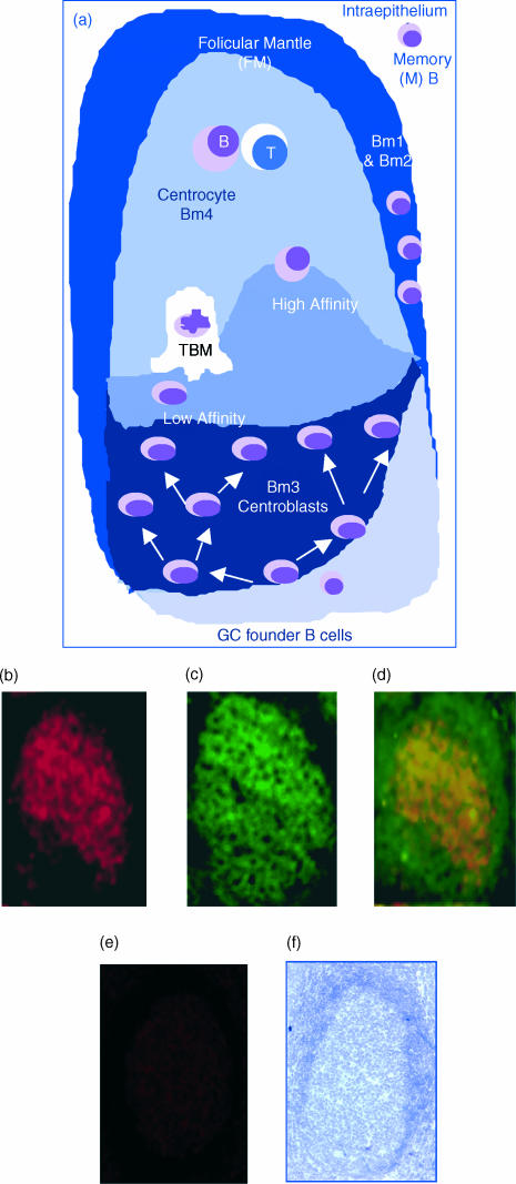
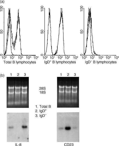
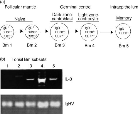
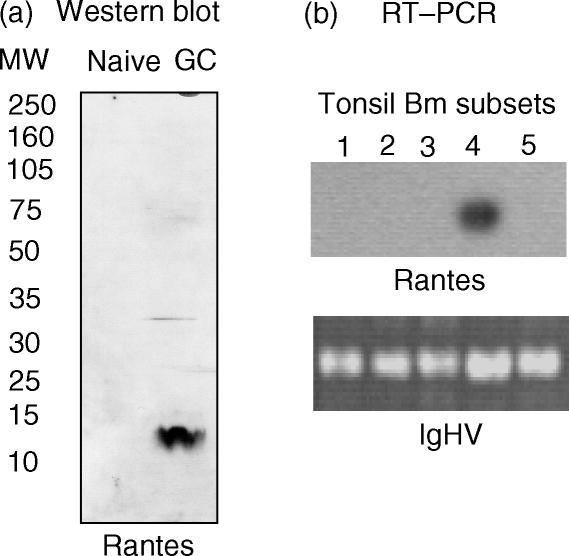
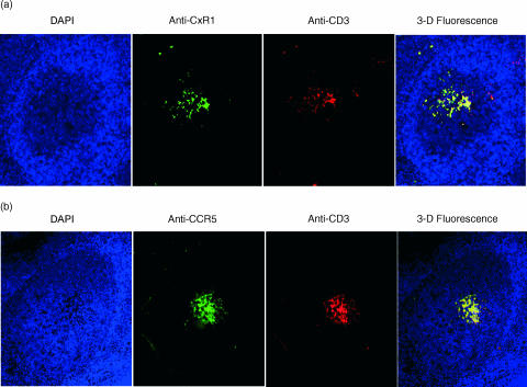
T-cell homing within germinal centres (GCs) is required for humoral B-cell responses. However, the mechanisms implicated in the recruitment of T cells into the GC are not completely understood. Here we show, by immunohistology, and Northern and Western blots, that in vivo human GC B lymphocytes can express CxC and CC chemokines. Moreover, B-cell subset-specific experiments reveal that interleukin (IL)-8 and regulated on activation, normal, T-cell expressed, and secreted (RANTES) are predominantly expressed by GC centroblast and centrocytes, suggesting that chemokine expression is essential at stages in which B-lymphocytes engage in active antigen-dependent interactions with T lymphocytes. In keeping with this hypothesis, we show that the T cells recruited into the GC correlatively express the receptors for IL-8 and RANTES. We propose that chemokine expression is a key B-cell function that facilitates T-lymphocyte recruitment into the GCs and supports cognate B-cell : T-cell encounters. Moreover, our data are consistent with the impaired homing of T cells to secondary lymphoid organs in mice that are either deficient in CC and CxC chemokines or their receptors.


Figures

Figure 1
Figure 1
Expression of interleukin (IL)-8 within germinal centres (GCs) by immunohistology. (a) A schematic representation of the GC reaction: an immunoglobulin M (IgM)-expressing GC founder migrates to the dark zone of a GC and undergoes clonal expansion and affinity maturation by somatic hypermutation. The mutated cells migrate to the basal light zone, where they are subjected to antigen-dependent selection. High-affinity (High) cells are selected and migrate to the apical light zone of GCs, where they encounter and present antigen (Ag) to T cells. Low affinity (Low) centroblasts undergo programmed cell death and are subsequently cleared by tingible body macrophages (TBM). (b) Reactivity of frozen tonsil (acetone-fixed) tissue sections to an affinity-pure anti-IL-8 immunoglobulin G (IgG). (c) B-cell identification by specific reactivity to an anti-CD20 monoclonal antibody (mAb). (d) Co-localization of the red and green fluorescence (anti-CD20), indicating the expression of IL-8 by CD20 B cells, as depicted by the yellow/orange merge. (e) Lack of reactivity to a non-immune IgG, used as a negative control. (f) Integrity of the tissue sections used, as determined by Gill's haematoxylin staining.
Figure 2
Figure 2
Differential expression of interleukin (IL)-8 and CD23 by immunoglobulin D+ (IgD+) and IgD B lymphocytes. (a) Flow cytometry profiles of pure IgD+ and IgD B-cell subpopulations. IgD cells co-localize with the mock isotype-matched control monoclonal antibody (mAb). (b) Northern blot analysis to determine the expression of IL-8 by total B cells and fluorescence-activated cell sorter (FACS) sorted IgD+ and IgD subsets. CD23 expression was used as control for the naïve IgD+ cells. RNA loading was routinely assessed by ethidium bromide staining of the agarose gels.
Figure 3
Figure 3
Predominant expression of interleukin (IL)-8 by centroblast and centrocyte B-lymphocyte subsets. (a) Phenotypic scheme of mature B-cell subsets: naive Bm1 and Bm2; germinal centre (GC) Bm3 and Bm4; and memory Bm5. (b) IL-8-specific reverse transcription–polymerase chain reaction (RT–PCR), using RNA from 500 cells of each Bm subset (Bm1 to Bm5). The numbers in the figure correspond to the respective Bm subsets. RT–PCR of the immunoglobulin heavy chain (IgHV) was used herein as control for the RNA content of each subset. The results are shown as ethidium bromide staining of electrophoresed RT–PCR samples.
Figure 4
Figure 4
Selective expression of regulated on activation, normal, T-cell expressed, and secreted (RANTES) by germinal centre (GC) B-cell subsets. (a) Western blot analysis depicting the selective expression of RANTES by IgD CD38+ GC B cells. (b) Southern blot experiment of a RANTES-specific reverse transcription–polymerase chain reaction (RT–PCR), using total RNA from 500 cells of each of the five mature B-cell subsets. The numbers in the figure correspond to the respective Bm1 to Bm5 subsets. Again, RT–PCR amplification of the immunoglobulin heavy chain (IgHV) was used as an internal control for the RNA content of each subset. The results are autoradiographs of the RANTES hybridization reaction and ethidium bromide gel staining of the IgHV RT–PCR results, respectively.
Figure 5
Figure 5
Concomitant germinal centre (GC) presence of T cells that express interleukin (IL)-8 and regulated on activation, normal, T-cell expressed, and secreted (RANTES) receptors. (a) Immunofluorescence detection of CxCR1-expressing T cells on tonsil tissue sections. (b) Histology and immunofluorescence for CCR5 expression by GC T cells. Starting from the left on both (a) and (b), the first panel depicts a histological section of a GC follicle revealed by the blue fluorescence of the 4′,6-diamidine-2-phenylindol dihydrochloride (DAPI) mount; the second panel displays the reactivity of either the anti-IL-8 receptor (CxCR1) or RANTES (CCR5) monoclonal antibodies (mAbs); the third panel reveals the detection of T cells by the reactivity to the anti-CD3 mAb; and the fourth panel is a three-dimensional merge of blue, green and red fluorescence.

References

    1. MacLennan ICM. Germinal centers. Annu Rev Immunol. 1994;12:117–39. - PubMed
    1. Kelsoe G. The germinal center: a crucible for lymphocyte selection. Semin Immunol. 1996;8:179–84. - PubMed
    1. Liu Y-J, Zhang J, Lane PJL, Chan EY-T, MacLennan ICM. Sites of specific B-cell activation in primary and secondary responses to T-cell-dependent and T-cell-independent antigens. Eur J Immunol. 1991;21:2951–62. - PubMed
    1. Jacob J, Kassir R, Kelsoe G. In situ studies of the primary immune response to [4-hydroxy-3-nitrophenyl] acetyl. I. The architecture and dynamics of responding cell populations. J Exp Med. 1991;173:1165–75. - PMC - PubMed
    1. Berek C, Berger A, Apel M. Maturation of immune responses in germinal centers. Cell. 1991;67:1121–9. - PubMed

Publication types

MeSH terms

LinkOut - more resources