. 2003 Dec;163(6):2173-8.
doi: 10.1016/S0002-9440(10)63574-2.

Hedgehog signaling regulates sebaceous gland development

Affiliations

Hedgehog signaling regulates sebaceous gland development

Mary Allen et al. Am J Pathol. 2003 Dec.

Abstract

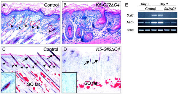
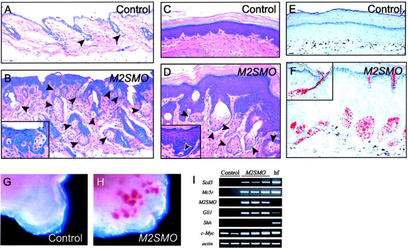
Epithelial progenitor cells in skin give rise to multiple lineages, comprising the hair follicle, an associated sebaceous gland, and overlying epidermis; however, the signals that regulate sebocyte development are poorly understood. We tested the potential involvement of the Hedgehog pathway in sebaceous gland development using transgenes designed to either block or stimulate Hedgehog signaling in cutaneous keratinocytes in vivo. Whereas inhibition of the Hedgehog pathway selectively suppressed sebocyte development, Hedgehog pathway activation led to a striking increase both in size and number of sebaceous glands. Remarkably, ectopic Hedgehog signaling also triggered the formation of sebaceous glands from footpad epidermis, in regions normally devoid of hair follicles and associated structures. These ectopic sebaceous glands expressed molecular markers of sebocyte differentiation and were functional, secreting their contents directly onto the skin's surface instead of into a hair canal. The Hedgehog pathway thus plays a key role in sebocyte cell fate decisions and is a potential target for treatment of skin disorders linked to abnormal sebaceous gland function, such as acne.


Figures

Figure 1.
Figure 1.
Dominant-negative inhibition of Gli activity blocks sebocyte development. A and B: H&E staining of skin from 7-day-old control and K5-Gli2ΔC4 transgenic mice. Clusters of sebocytes are seen associated within the superficial portion of control hair follicles (dashed red lines and arrowheads in A, but not in aberrant hair follicles arising in transgenic skin in B). C and D: Oil Red O staining for lipids. Note the presence of large, amorphous masses of adipocytes staining in subcutaneous fat layer (SQ fat) in both the control (C) and transgenic (D) skin sections. Staining of sebaceous glands is limited to control skin (inset and arrowheads in C). Despite the reduced length of follicles in K5-Gli2ΔC4 mice (compare B and D with A and C), the transgenic hair follicles express multiple markers for hair follicle lineages (in preparation) and assemble abortive, pigmented hair shafts (arrows in D). E: Semiquantitative RT-PCR for sebocyte markers reveals up-regulation of Scd3 and Mc5r in control skin between postnatal day 1 and day 9, reflecting expansion of the sebocyte lineage during this time. Expression of transcripts encoding sebocyte markers in skin from day 9 K5-Gli2ΔC4 samples (Gli2ΔC4) was similar to that of day 1 control samples.
Figure 2.
Figure 2.
Enhanced activation of Shh signaling in keratinocytes leads to formation of hyperplastic and ectopic sebaceous glands. A and B: H&E staining of control and M2SMO-expressing dorsal mouse skin at 6.5 months of age. Note small size of normal sebaceous glands in control skin (arrowheads in A) compared with increased number and size of sebaceous glands in transgenic skin (arrowheads in B). In addition to glandular structures within the dermis, individual sebocytes or small aggregates are also found ectopically within the epidermis (inset in B). C and D: H&E staining of volar paw skin from control and transgenic mice at 13 months of age. Note thickened epidermis and cornified cell layers typically seen in skin from this region, with a conspicuous absence of hair follicles and sebaceous glands (C). Ectopic sebaceous glands (arrowheads in D) develop from volar epidermis of mice expressing M2SMO. Sebaceous gland development is initiated at the base of epidermal downgrowths (inset in D). E and F: Oil Red O staining for lipids in control and transgenic volar skin. As expected, no staining is detected in control skin in E, whereas multiple sebocytes are stained in transgenic mouse skin in F. Many of the ectopic sebaceous glands secrete Oil Red O-positive material directly onto the skin’s surface (see inset). G and H: Whole-mount analysis of footpads from control and transgenic mice. A large number of Oil Red O-positive sebaceous glands are detected in transgenic (H) but not control (G) footpad. I: Semiquantitative RT-PCR analysis showing expression of sebocyte markers (Scd3, Mc5r) in volar skin from M2SMO-expressing transgenic mice (M2SMO) and control skin containing hair follicles (hf), but not in control volar skin. Transgene-specific primers confirm expression of M2SMO in transgenic samples, which also contain elevated levels of the Shh target gene Gli1.

References

    1. Fuchs E, Merrill BJ, Jamora C, DasGupta R: At the roots of a never-ending cycle. Dev Cell 2001, 1:13-25 - PubMed
    1. Niemann C, Watt FM: Designer skin: lineage commitment in postnatal epidermis. Trends Cell Biol 2002, 12:185-192 - PubMed
    1. Byrne C, Hardman M, Nield K: Covering the limb: formation of the integument. J Anat 2003, 202:113-123 - PMC - PubMed
    1. Hardy MH: The secret life of the hair follicle. Trends Genet 1992, 8:55-61 - PubMed
    1. Cotsarelis G, Sun TT, Lavker RM: Label-retaining cells reside in the bulge area of pilosebaceous unit: implications for follicular stem cells, hair cycle, and skin carcinogenesis. Cell 1990, 61:1329-1337 - PubMed

Publication types

LinkOut - more resources